Cat. #151755
C10 Colorectal Cell Line
Cat. #: 151755
Unit size: 1x10^6 cells / vial
Organism: Human
Tissue: Colon
Disease: Cancer
Model: Tumour line
£575.00
This fee is applicable only for non-profit organisations. If you are a for-profit organisation or a researcher working on commercially-sponsored academic research, you will need to contact our licensing team for a commercial use license.
Contributor
Inventor: Walter Bodmer
Institute: University of Oxford
Tool Details
*FOR RESEARCH USE ONLY (for other uses, please contact the licensing team)
- Name: C10 Colorectal Cell Line
- Cancer type: Colorectal cancer
- Cancers detailed: Colorectal;Dukes' stage B;Human colorectal adenocarcinoma;Moderately well differentiated
- Research fields: Cancer;Drug development
- Organism: Human
- Tissue: Colon
- Disease: Cancer
- Model: Tumour line
- Conditional: Yes
- Description: The C10 cell line was established from a 71-year old male patient with moderately well differentiated adenocarcinoma of the descending colon classified as Dukes' stage B. C10 harbours a well-characterised TP53 point mutation located in exon 7, codon 245, involving a G-to-A nucleotide substitution that results in a glycine-to-serine amino acid change.
- Production details: This colorectal cell line was established from surgically resected colorectal adenocarcinomas.
- Cellosaurus id: CVCL_5245
Handling
- Format: Frozen
- Growth medium: Iscove's Modified Dulbecco's Medium, + 10% Foetal Bovine Serum (FBS) + 2mM Glutamine
- Temperature: 37° C
- Atmosphere: 5% CO2
- Unit size: 1x10^6 cells / vial
- Shipping conditions: Dry ice
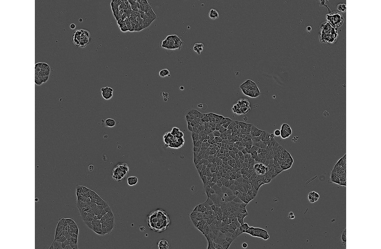
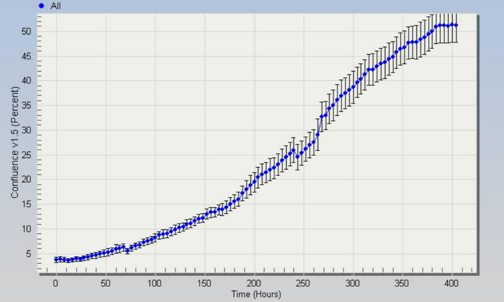
- Subculture routine: Split sub-confluent cultures (70-80%) 1:3 to 1:6 i.e. seeding at 2-4x10,000 cells/cm² using 0.05% trypsin or trypsin/EDTA; 5% CO₂; 37°C. C10 cells grow very slowly; following resuscitation or subculture the cells take at least 48 hours to re-attach. Cells should be left without disturbance during this time to facilitate adhesion. Centrifugation of the cells (100g x 5 min) at resuscitation to remove DMSO improves the establishment of a viable culture. Once attached, the cells grow in discrete islands and use of trypsin or trypsin/EDTA to subculture the cells (even without knocking the flask) yields large clumps. Further disaggregation may be achieved by repeatedly pipetting the cells.
References
- Bracht et al. 2010. Br J Cancer. 103(3):340-346. PMID: 20606684.
- Liu et al. 2006. Proc Natl Acad Sci U S A. 103(4):976-981. PMID: 16418264.
- Rowan et al. 2000. Proc Natl Acad Sci U S A. 97(7):3352-3357. PMID: 10737795.
- Efstathiou et al. 1999. Proc Natl Acad Sci U S A. 96(5):2316-2321. PMID: 10051639.
- Browning et al. 1993. Proc Natl Acad Sci U S A. 90(7):2842-2845. PMID: 8464898.

![Anti-CAR Whitlow Linker [1C3C3]](https://cancertools.org/wp-content/uploads/Figure-6-Kimble-et-al.-J-Immunother-Cancer-2025-300x322.jpg 300w, https://cancertools.org/wp-content/uploads/Figure-6-Kimble-et-al.-J-Immunother-Cancer-2025-280x300.jpg 280w, https://cancertools.org/wp-content/uploads/Figure-6-Kimble-et-al.-J-Immunother-Cancer-2025-954x1024.jpg 954w, https://cancertools.org/wp-content/uploads/Figure-6-Kimble-et-al.-J-Immunother-Cancer-2025-768x824.jpg 768w, https://cancertools.org/wp-content/uploads/Figure-6-Kimble-et-al.-J-Immunother-Cancer-2025.jpg 1193w)