Brainbow antibodies: from Dawen Cai’s lab to researchers worldwide

Parvalbumin expressing neurons labelled by AAV-Brainbow and stained with Brainbow 3 XFP antibodies against mTFP, mCherry, TagRFP, and EGFP. Image credit: Dawen Cai

Originally developed to visualise complex neural circuits, Brainbow fluorescent protein (XFP) antibodies have become a powerful tool for tracking individual cells, amplifying multicolour signals, and enabling discoveries across tissues and organisms. We spoke with Prof. Dawen Cai from the University of Michigan, the inventor of these polyclonal antibodies against XFPs originated from very distinct spieces, to hear why he developed them and the impact they are having in both neuroscience and cancer research. By making these antibodies widely available, Prof. Cai is helping researchers to build on proven innovation, focus their efforts on new discoveries and accelerating progress across multiple fields.

Dawen Cai, PhD.

Dawen Cai, PhD. University of Michigan

Seeing the brain clearly

Neuroscience is rapidly advancing through new antibodies and imaging technologies, allowing researchers to visualise the brain in remarkable detail. These insights are reshaping our understanding of how the brain develops, functions, and fails in disease.

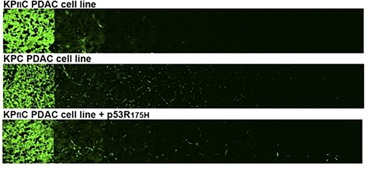
For the cancer research community, this progress is increasingly relevant. Tumours often interact with surrounding tissue, including neural networks in the brain and peripheral nervous system. For example, glioblastoma research has shown that tumours do not invade the brain randomly and instead, hijacks the brain’s own neuronal mechanisms through malignant crosstalk, transforming normal brain function into an ecosystem for tumour progression (1). In brain cancers and in cancers that invade or communicate with nerves, understanding how malignant cells interact with neurons and their microenvironment requires imaging tools that can resolve neuronal structure and functions within living tissue.

Despite advances, a fundamental technical challenge persists: how can we visually distinguish individual neurons and cancer cells when they are packed together in dense, anatomically complex tissue? This limitation affects both basic neuroscience and translational cancer biology, where mapping cellular interactions at scale is essential.

The limits of traditional neural labelling

Traditional labelling methods, such as Golgi staining or dye injection, can reveal the morphology of a handful of neurons at a time, but never provide the complete picture. Fluorescent protein approaches have expanded what researchers can see, but still often result in weak signals, poor labelling of fine structures, and limited colour diversity. As a result, many studies have been forced to choose between imaging a small number of neurons clearly or many neurons indistinctly — a trade-off that has slowed progress in understanding both brain circuitry and disease, including cancer-related changes in the nervous system.

A major advancement came in 2007 with the introduction of Brainbow technology, developed by Jeff Lichtman and Josh Sanes at Harvard. By engineering transgenic mice to express different combinations of fluorescent proteins, researchers could label individual neurons in ~ 100 distinct colour combinations. This transformed the study of neural connectivity and made large-scale neural circuit mapping possible (2).

However, during Prof. Cai’s time at Harvard, he and his colleagues became aware of practical limitations that were preventing Brainbow from reaching its full potential. In particular:

  • The original fluorescent proteins were prone to photoinstability and often produced weak or inconsistent signals.
  • Because the proteins were cytoplasmic, they accumulated in neuronal cell bodies, failing to adequately label delicate axons and dendrites where critical connectivity information resides.
  • The non-recombinant state expressed red fluorescent protein (RFP), producing a dominant “default” red colour that reduces true diversity across labelled neurons.
  • The fluorescent proteins shared too much sequence homology to allow the design of specific immunostaining antibodies, making it impossible to recover signal once native fluorescence was lost during fixation, sectioning or tissue clearing.

To overcome these limitations, Prof. Cai and his team developed the Brainbow XFP antibodies, creating a new generation of tools that make multicolour neural imaging brighter, more reliable, and more broadly applicable.

The Brainbow antibodies solve a fundamental bottleneck in multicolor fluorescent labeling: they provide a robust, species-agnostic way to amplify and recover the fluorescent signal after tissue fixation or processing, which is essential for any multicolor labeling experiment to be reliably quantified and compared across labs.

Dawen Cai, PhD
Cholenergic neurons labelled by AAV-Brainbow and stained with Brainbow XFP antibodies against mTFP, mCherry, TagRFP, and EGFP. Image credit, Dr. Douglas Roossien.

Cholenergic neurons labelled by AAV-Brainbow and stained with Brainbow XFP antibodies against mTFP, mCherry, TagRFP, and EGFP. Image credit: Dr. Douglas Roossien.

From Brainbow 1 to Brainbow 3: designing better fluorophores

Prof. Cai developed Brainbow 3 with the selection of improved XFPs that were brighter, more photostable, and less prone to aggregation than those used in earlier versions. As a result, the original fluorophores were replaced with mOrange2 (from coral), EGFP (from jellyfish), and mKate2 (from sea anemone), significantly improving signal quality and stability.

The team also engineered farnesylated derivatives of these proteins, so they localised to the cell membrane rather than accumulating in the cell body. This adaptation dramatically improved labelling of fine neuronal structures, including axons and dendrites — features critical for mapping neural connectivity.

Building on this, Prof. Cai’s group expanded the colour palette further through new transgene designs, Brainbow 3.1, 3.2, and AAV-Brainbow. By incorporating a broader range of XFPs, the system supported more distinct colours, eliminated the dominant “default red” problem, and enabled clearer discrimination between neighbouring neurons. Regulatory elements, including “stop” cassettes, were also introduced to prevent default expression of a single fluorophore and promote a more balanced colour distribution across labelled cells.

The key challenge: achieving true antibody specificity

Developing Brainbow 3 was not straightforward. Fluorescent proteins belong to a relatively conserved family, meaning that antibodies raised against closely related proteins often cross-react – a problem that would undermine multicolour labelling strategy. For immunostaining to work alongside Brainbow, each antibody needed to cleanly distinguish one fluorophore from all others.

The solution was strategic fluorescent protein selection. By choosing proteins from diverse evolutionary lineages – including coral, jellyfish, and sea anemones – the team maximised sequence divergence and antigenic distance. This enabled the generation of highly specific antibodies with minimal cross-reactivity. Ultimately, the panel comprised eight spectrally and antigenic distinct fluorescent proteins: mTFP1, EGFP, mNeonGreen, TagYFP, PhiYFP, mKusabiraOrange2, TagRFP, and mCherry.

Making the antibodies work in practice

A further challenge was ensuring the antibodies performed reliably across real-world experimental settings. Brainbow is used in transgenic mice, via AAV vectors, in zebrafish and Drosophila, and across a wide range of tissue processing workflows. To maximise compatibility, Prof. Cai’s team generated <strong>custom polyclonal antibodies in multiple host species — including chicken, rabbit, rat, and guinea pig — giving researchers more flexibility in choosing a broad range of secondary antibodies.

These antibodies were validated in cells and mice to confirm that they amplified signals without compromising colour diversity. Crucially, the team demonstrated that antibody amplification enhanced signal strength while preserving the spectral distinctions required for multicolour analysis — a result that underpinned their broad adoption (3).

Transgenic Bitbow Drosophila larval brain. The novel Bitbow design integrates five antigenic distinct FPs (mAmetrine, mTFP1, mNeonGreen, mKusabiraOrange2, and tdKatushka2) that are targeted to the nucleus, Golgi aparatus, and cytoplasmic membrane. After Flp induced random recombination, the 15-bit binary subcellular spectral barcodes are stained with the Brainbow XFP antibodies and utilised for neuron lineage tracing. Image credit, Dr. Ye Li

Transgenic Bitbow Drosophila larval brain. The novel Bitbow design integrates five antigenic distinct FPs (mAmetrine, mTFP1, mNeonGreen, mKusabiraOrange2, and tdKatushka2) that are targeted to the nucleus, Golgi aparatus, and cytoplasmic membrane. After Flp induced random recombination, the 15-bit binary subcellular spectral barcodes are stained with the Brainbow XFP antibodies and utilised for neuron lineage tracing. Image credit: Dr. Ye Li

The outcome: a robust, versatile Brainbow XFP antibody toolkit

This improved antibody panel became the critical enabling technology. It transformed the original Brainbow from a technique reliant solely on native fluorescence into a robust, versatile immunostaining toolkit that performs reliably across fixation, tissue clearing, expansion microscopy, and a wide range of experimental settings.

For any researcher working with fluorescent protein-based cell labelling — whether in neuroscience, cancer biology, or developmental biology — the Brainbow antibodies transform a technically fragile approach into a dependable, standardised toolkit.

Dawen Cai, PhD

From one lab to the world: sharing the Brainbow for faster discovery

Accessing research tools developed in other academic labs has normally involved lengthy email exchanges, complex material transfer agreements, and unpredictable shipping timelines — all of which can slow scientific progress. Through the non-profit research tool platform CancerTools, access to <strong>Brainbow XFP antibodies is now streamlined and reliable, reducing these practical barriers and enabling researchers to adopt validated tools more quickly.

As Prof. Cai explained, the Brainbow XFP antibodies were initially developed to solve a technical challenge within his own laboratory, but it soon became clear that this problem was universal. By making these antibodies available through CancerTools, laboratories worldwide can access a proven, well-characterised solution without having to recreate and optimise from scratch, an approach that reflects our shared mission with Cancer Research UK.

By enabling wide access to Brainbow XFP antibodies, Prof. Cai has helped cultivate a culture of openness and collaboration, ensuring that his work continues to empower researchers across neuroscience and cancer biology.

Personally, it has reinforced my belief that the most impactful thing a methodologist can do is not just develop tools but make them accessible. The barrier to adoption matters enormously. A brilliant tool that sits in one lab is worth far less than a good tool that hundreds of labs can pick up and use.

Dawen Cai, PhD

From brain circuits to cancer discovery

In neuroscience, Brainbow antibodies continue to support high-resolution circuit mapping by preserving signal through fixation, tissue clearing, and advanced imaging — capabilities that now extend beyond the brain.

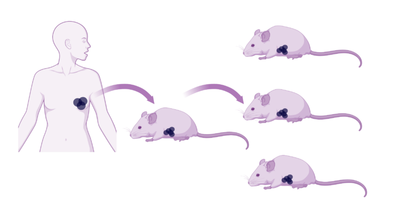
What has been particularly striking is how the Brainbow framework has inspired new approaches in cancer biology. A leading example is the Cancer Rainbow (Crainbow) mouse model developed by the Snyder laboratory at Duke University. Building directly on Brainbow’s principle of combinatorial fluorescent barcoding, the team paired specific oncogenic mutations, such as β-catenin (Ctnnb1), with distinct fluorescent proteins, allowing visualisation of clonal expansion in living tissue (4). Their work revealed how developmental timing and microenvironmental signals such as oncogenic RSPO3 can drive the emergence and spread of premalignant clones in adult intestines, helping explain tumour heterogeneity and early cancer evolution.

While Brainbow antibodies were not the primary tool used in Crainbow, the multicolour fluorescent protein principles pioneered by Brainbow made experiments like this possible. Equally, antibody-based signal amplification remains essential whenever robust detection is required in fixed or processed tissues. This is a powerful example of how one foundational technology can seed entirely new innovations in cancer research.

It has been deeply rewarding to see the Brainbow antibodies adopted by labs working on questions I would never have imagined when we first developed them. The moment that really crystallized this for me was learning about the Crainbow work at Duke — seeing how the principles of multicolor fluorescent barcoding had been adapted to directly visualise the spread of oncogenic mutations in the gut, and how that led to the surprising finding that colorectal cancer may be seeded during early postnatal development. That is the kind of impact that makes tool development feel truly worthwhile — when a technology you built opens a door into a completely new area of biology.

Dawen Cai, PhD

Brainbow in the years ahead

Brainbow antibodies are more than a neuroscience tool – they form a general-purpose toolkit for experiments that rely on stochastic multicolour fluorescent labelling. For cancer researchers, they represent a powerful opportunity to visualise clonal evolution, tumour heterogeneity, and interactions with the tumour microenvironment, opening new approaches to study how cancers arise, diversify, and progress at the cellular level.

The development and widespread adoption of Brainbow 3 by Prof. Dawen Cai exemplify how thoughtfully designed research tools can shape the future of science. By advancing neuroscience while enabling innovation in cancer biology, his work provides a foundation that researchers can build on — supporting new insights and discoveries for years to come.

Parvalbumin expressing neurons labelled by AAV-Brainbow and stained with Brainbow 3 XFP antibodies against mTFP, mCherry, TagRFP, and EGFP. Image credit: Dawen Cai

Parvalbumin expressing neurons labelled by AAV-Brainbow and stained with Brainbow 3 XFP antibodies against mTFP, mCherry, TagRFP, and EGFP. Image credit: Dawen Cai

Explore the Brainbow antibodies to power your next discovery

References:

  1. Venkataramani, V., et al. 2022. Cell. 185(16), 2899-2917. PMID: 35914528
  2. Livet, J., et al. 2007. Nature. 450, 56-62. PMID: 17972876
  3. Cai, D., et al. 2013. Nature Methods. 10, 540-547. PMID: 23817127
  4. Boone, P.G., et al. 2019. Nature Communications. 10, 5490.PMID: 31792216

How are researchers using the KPC cell line to answer the toughest questions in pancreatic cancer?

KPC Cell Line (C57/BL6 genetic background) cell sheet monolayer
Prof. Jennifer Morton, Cancer Research UK Scotland Institute

Prof. Jennifer Morton, Cancer Research UK Scotland Institute

In our previous article, we introduced the origins of the KPC cell line, developed by Professor Jennifer Morton at Cancer Research UK’s Scotland Institute. Here we focus on what matters most: how the KPC line is being used to answer some of the toughest questions in pancreatic cancer. From modelling metastasis and tumour-immune interactions to testing therapeutic strategies in immunocompetent systems, the KPC cell line offers a transplantable, genetically defined model that combines biological relevance with experimental flexibility. The studies highlighted below show why KPC has become a preferred platform for researchers who need to move efficiently between in vitro perturbations and immunocompetent in vivo validation.

From foundational models to practical experimental tools

The original KPC genetically engineered mouse model transformed pancreatic cancer research by revealing key drivers of tumour progression, stromal remodelling, and immune suppression. Today, scientists across the world are building on these foundational insights through the transplantable KPC-derived cell line established by Prof. Jennifer Morton at the Cancer Research UK Scotland Institute, which allows these key biological questions to be explored with greater experimental flexibility.

The KPC model enables defined genetic perturbation and functional analysis; here, PTEN loss increases macropinocytic uptake of fluorescent dextran (red) (adapted from Michalopoulou et al.).  

By separating tumour genetics from tumour initiation, a key limitation of spontaneous models, the KPC cell line allows researchers to control timing, introduce defined perturbations, and apply treatments reproducibly, while still working in immunocompetent hosts. Compared with spontaneous models, they enable tumour genetics, timing, and treatment to be manipulated independently, supporting experiments that would otherwise require lengthy breeding strategies.

What began as a tool to study tumour biology in a more controlled way has since evolved into a platform for tackling increasingly complex questions, from adaptive resistance to treatment delivery and tumour–immune dynamics. Here is why researchers are extending the original vision of the model in practice.

Using the KPC line to study tumour adaptation

Tumour adaptation is a core reason why many targeted therapies fail; understanding it can guide more effective combinations.

A central challenge in pancreatic cancer research is understanding how tumour cells adapt when key signalling or metabolic pathways are disrupted, particularly as such adaptation often underlies therapeutic resistance. In spontaneous models, introducing additional genetic changes or systematically testing resistance mechanisms often requires long timelines. Several groups have therefore chosen the KPC cell line as a genetically defined but tractable baseline for studying tumour adaptation under selective pressure, such as pathway inhibition and nutrient stress.

At the Cancer Research UK Scotland Institute (formerly the Beatson Institute), Michalopoulou and colleagues compared KPC and KCPTEN pancreatic cancer cell lines derived from genetically engineered mouse models to examine how PTEN loss reshapes nutrient uptake and influences response to mTOR inhibition. By combining in vitro metabolic and uptake assays with mechanistic perturbations, the team showed that PTEN loss enhances macropinocytosis, a process by which cells engulf extracellular material, allowing tumour cells to scavenge extracellular protein and sustain growth despite mTOR inhibition. This mechanism could be pharmacologically targeted by inhibiting lysosomal degradation, revealing a therapeutic vulnerability.

Related studies from multiple universities have used KPC-based perturbation models to examine tumour adaptation under defined genetic or treatment pressure, including circadian clock disruption shaping tumour growth in vivo and chemotherapy response (Schwartz et al.). Orthotopic KRAS-driven syngeneic models, in which tumour cells are implanted into the pancreas of immunocompetent genetically matched mice, have likewise been used to interrogate tumour–stroma signalling under defined perturbation. For example, genetic and pharmacological targeting of the ACSL3–PAI-1 axis reshaped fibrosis and altered immune cell infiltration in vivo (Sebastiano et al.). Disrupting mechanosensing pathways has shown how the physical properties of the tumour environment can influence fibroblast behaviour and tumour growth in vivo (Romac et al.).

Taken together, these studies show how the KPC cell line provides a controlled starting point for layering defined genetic or pharmacological perturbations and tracking tumour behaviour over time in vivo. By enabling rapid genetic engineering, quantitative metabolic and uptake assays in vitro, and validation in vivo, including syngeneic contexts, this approach allows researchers to interrogate how pancreatic cancer cells adapt under selective pressure without the constraints of spontaneous tumour development.

While the work above focuses on tumour-intrinsic mechanisms of adaptation, the same experimental flexibility can allow you to ask how treatment context itself—such as timing, delivery, or local intervention—shapes tumour response, helping you design experiments with improved translational impact.

Using KPC to test therapies in realistic in vivo contexts

How and when you deliver a therapy can be as important as the therapy itself, and transplant models let you study that explicitly.

Many clinically relevant questions in pancreatic cancer depend not only on which therapy is used, but also on when, where, and how it is delivered – factors that can substantially influence treatment outcome. Spontaneous tumour models can make such questions difficult to address, as tumour onset and progression are less predictable. By contrast, the KPC cell line allows tumours to be generated with defined timing and location, enabling researchers to align tumour growth more directly with treatment delivery and experimental intervention.

At the University of Glasgow, Falcone and colleagues used the KPC line to generate subcutaneous tumours in immunocompetent mice and examined how dietary serine and glycine availability influences radiotherapy response. By controlling both diet and radiation exposure, the group showed that amino-acid restriction increased response to radiotherapy. Rather than focusing on immune mechanisms directly, the study highlighted how systemic context, such as diet, can shape tumour response in ways not captured by standard in vitro assays.

Additional studies have applied orthotopic KPC tumour models to reconstruct clinically relevant intervention scenarios under controlled conditions, including local ablative therapy using irreversible electroporation (Woeste et al.).

The orthotopic KPC model enables testing of combination therapies in immunocompetent mice; here, β-glucan combined with irreversible electroporation reduces tumour burden (Woeste et al.).

In these settings, researchers use KPC-based transplantation models not to discover new driver mutations, but to model treatment delivery and response under controlled conditions. By combining syngeneic or orthotopic tumour implantation with defined local or systemic interventions and longitudinal assessment, KPC models allow researchers to examine how treatment design, local control, and host context influence tumour progression in immunocompetent hosts. This level of experimental control enables interrogation of tumour behaviours, such as host immune responses and tumour microenvironment dynamics, that depend on interactions with an intact host environment and are not readily captured in vitro.

If your work involves testing treatment timing, delivery, or combination strategies in vivo, the KPC cell line provides a way to do so within a defined genetic background and immune-competent setting, without the variability and timelines associated with spontaneous models.

Using KPC to model immune-dependent tumour behaviour in vivo

Interactions between tumour cells and the immune system are fundamentally different in vivo than in vitro, and models that capture this are essential for immunotherapy research.

Some tumour behaviours in pancreatic cancer are fully revealed when tumour cells interact with an intact immune system in vivo. To address biological questions that cannot be resolved in vitro, several groups have applied the KPC cell line as syngeneic allografts in immunocompetent hosts.

At the University of Glasgow, Newman and colleagues used parental and IDO1-engineered KPC cell lines to examine how immune-derived interferon-γ influences tumour metabolism in vivo. By implanting these lines into immunocompetent mice, the team showed that interferon-γ induces high IDO1 expression, activating the kynurenine pathway and enabling tumour cells to channel tryptophan metabolism into nucleotide biosynthesis. This immune-conditioned metabolic dependency was not apparent under standard culture conditions and emerged in the presence of host-derived signals.

Beyond tumour-intrinsic metabolic adaptation, KPC-derived tumours have also been used to interrogate immune behaviour that emerges only in immunocompetent hosts. Orthotopic KPC models have been used to demonstrate trained innate immunity within the pancreatic tumour microenvironment (Geller et al.), to show that local ablative therapy can further remodel myeloid activation states in vivo (Woeste et al.), and to reveal that microbiota-derived metabolites can influence chemotherapy response via neutrophil-derived myeloperoxidase (Tintelnot et al.). Together, these studies illustrate how host context and treatment exposure reshape tumour–immune interactions in ways not apparent under standard culture conditions and that depend on intact immune competence.

Orthotopic KPC-derived tumours in immunocompetent C57BL/6 mice, illustrating in vivo treatment-associated changes assessed by histology (Tintelnot et al.).

In these examples, the KPC cell line bridges reductionist systems and physiological complexity, allowing you to examine tumour-intrinsic processes within an immune-competent environment, while maintaining precise control over tumour genetics and timing.

Interpreting results and experimental caveats – why careful model interpretation matters!

As with any transplantable system, careful interpretation of data generated using the KPC cell line is essential. Studies have shown that additional genetic modifications, such as reporter gene expression, can alter tumour–immune interactions in immunocompetent hosts. For example, luciferase-expressing KPC tumours, in which a foreign reporter protein is introduced, have been shown to provoke immune responses that influence tumour growth dynamics in immunocompetent hosts (Ferrari et al.).

More broadly, the KPC cell line is most powerful when used alongside complementary models: it offers speed, experimental control, and accessibility that are difficult to achieve with spontaneous tumours, while necessarily sacrificing aspects of tumour initiation and long-term evolution. For researchers, this translates into faster hypothesis testing and greater experimental precision, with the option to validate findings in more complex models when needed.

Looking ahead

Across these examples, a clear pattern emerges: researchers are applying the KPC cell line not as simplified stand-ins, but as enabling platforms for asking nuanced experimental questions.

As research priorities increasingly focus on combination therapies, adaptive resistance, and tumour–microenvironment interactions, the KPC cell line provides a practical and experimentally accessible system for interrogating these challenges under controlled conditions. Its ability to bridge defined in vitro perturbations with immunocompetent in vivo validation positions it as a foremost platform for shaping preclinical pancreatic cancer research.

Explore the KPC cell line and see how this model can accelerate your next pancreatic cancer study

References

Falcone M et al. 2022. British Journal of Cancer. 127:1773–1786. PMID: 36115879.  

Ferrari DP et al. 2024. Sci Rep. 14(1):13602. PMID: 38866899. 

Geller A et al. 2022. Nature Communications. 13(1):759. PMID: 35140221.  

Michalopoulou E et al. 2020. Cell Reports. 30(8):2729–2742.e4. PMID: 32101748.  

Newman AC et al. 2021. Mol Cell. 81(11):2290–2302.e7. PMID: 33831358.  

Romac JMJ et al. 2025. JCI Insight. 10(23):e196280. PMID: 41100488.  

Schwartz PB et al. 2023. PLoS Genet 19(6): e1010770. PMID: 37262074.  

Sebastiano MR et al. 2020. Sci. Adv. 6(44):eabb9200. PMID: 33127675.  

Tintelnot J et al. 2023. Nature. 615(7950):168-174. PMID: 36813961 

Woeste MR et al. 2023. J Immunother Cancer. 11(4):e006221. PMID: 37072351. 

Dissecting the Bladder Tumour Microenvironment with MB49

Introduction

Immunotherapy has started to transform bladder cancer treatment, but durable responses remain the exception rather than the rule. Understanding why requires models that capture the tumour microenvironment, not just tumour growth. This blog explores how the MB49 syngeneic bladder cancer model – across subcutaneous, orthotopic, MB49-luc, and metastatic formats – dissects myeloid-dominated “cold” TMEs, neutrophil-driven immune escape, BCG response, and PD-L1-dependent checkpoint timing.

Bladder cancer is clinically heterogeneous and notoriously difficult to treat once it progresses to muscle-invasive disease. Immune checkpoint blockade and intravesical immunotherapy have transformed parts of the treatment landscape. Yet durable responses remain limited to a subset of patients. The field therefore relies on robust preclinical models that recapitulate tumour-immune interactions and reveal why therapies succeed or fail.

Among these, MB49 has emerged as one of the most reliable and informative models. Originally derived from chemically induced urothelial carcinoma arising in C57BL/ICRF-a(t) mouse bladder epithelium (1), MB49 has become a workhorse for studying the bladder tumour microenvironment (TME) and its manipulation by immunotherapy.

light microscope image of MB49 in culture
Image: Light microscope image of MB49 cells

What makes MB49 so valuable for bladder cancer research?

Its value lies in the combination of syngeneity and biological relevance (2–6):

  • Reliable growth in immunocompetent models: MB49 grows readily in C57BL/6‑background mice, allowing studies in the context of intact host immunity (2–6).
  • Epithelial, urothelial origin: The line is derived from chemically induced urothelial carcinoma and therefore recapitulates the epithelial nature of human bladder cancer (1).
  • Clinically relevant antigen and checkpoint expression: MB49 expresses key molecules such as PD‑L1 and chemokines that are central to current immunotherapy strategies (1,4–6).
  • Myeloid‑dominated, “cold” tumour microenvironment: Orthotopic MB49 tumours show a TME rich in suppressive myeloid cells with comparatively sparse T‑cell infiltration, closely resembling many human bladder tumours (2,4–6).

This “immunologically cold” phenotype mirrors a large fraction of human bladder tumours. MB49 therefore excels at dissecting immune evasion, myeloid–tumour crosstalk, and heterogeneous immunotherapy responses (2,4–7). Over the past two decades, MB49 underpins some of the clearest mechanistic insights into how the bladder TME becomes hostile to anti-tumour immunity, and how that state might be reversed. This impact is reinforced by MB49’s flexibility across multiple in vivo formats.

Choosing the right MB49 model

Researchers use MB49 in four main in vivo formats, each offering a slightly different window onto the TME and different practical advantages; subcutaneous, orthotopic, MB49-luc, and metastatic models (Table 1).

Subcutaneous MB49 tumours grow quickly and reproducibly, enabling clean readouts of tumour volume alongside rich TME profiling, ideal for dissecting innate pathways, macrophage polarisation, and checkpoint‑modulating therapies (3).

Orthotopic MB49 tumours, generated by intravesical instillation into pre‑injured bladders, add bladder‑specific factors such as urothelium, urine contact, and organ‑specific immune recruitment. This makes orthotopic models particularly powerful for intravesical BCG, gene therapy, and bladder‑targeted immunotherapy studies (3).

MB49-luc, a luciferase-tagged derivative of parental MB49 instilled orthotopically, enables longitudinal imaging and real-time TME tracking. Perfect for studying PD-L1 kinetics and therapy timing (4).

Metastatic MB49 models, typically generated via intravenous injection, extend investigations to distant immune niches, commonly the lung. These models excel at studying systemic immunity and dissemination (3).

Model type Setup Best Use Case Advantages Disadvantages
Subcutaneous MB49 cells injected under the skin (flank or hind leg) Rapid efficacy and dosing studies; general TME and immune‑modulation; combo immunotherapy Technically simple; highly reproducible; easy tumour measurement and tissue access TME is skin/subcutis, not bladder; lacks urine exposure and bladder‑specific stromal context
Orthotopic MB49 cells instilled into the bladder via catheter after urothelial pre‑injury Bladder‑specific TME, intravesical BCG or gene therapy, modelling NMIBC progression Anatomically and physiologically closest to human bladder cancer; native bladder microenvironments Technically demanding; variable take; tumour monitoring often needs imaging or defined endpoints
MB49-luc (orthotopic) Luciferase-tagged MB49 cells instilled intravesically Longitudinal TME tracking; PD-L1 kinetics; intravesical therapy timing Non-invasive bioluminescence imaging; temporal immune dynamics Requires imaging equipment; luciferase signal variability
Metastatic MB49 cells delivered IV (tail vein) Metastatic spread, lung TME, systemic immunotherapy and adoptive cell therapy Models dissemination and distant‑organ TME; suited to systemic therapy readouts Primary tumour not in bladder; metastatic organ context differs; quantification more laborious

Table 1: MB49 in vivo model formats – use cases and recommendations (3,4).

MB49 in action: neutrophils as architects of immune escape

One of the clearest examples of MB49’s translational impact comes from its role in understanding the function of neutrophils in bladder cancer. Clinically, high neutrophil infiltration has long been associated with poor prognosis (7), but the causality and mechanism were unclear.

Using orthotopic MB49 models, Jing et al. demonstrated that tumour-derived CXCL1, a major murine functional analogue of human IL-8, drives robust neutrophil recruitment into the bladder TME (7). Once present, these neutrophils secrete hepatocyte growth factor (HGF). This activates MET signalling in tumour cells and amplifies CXCL1 production. This results in a self-reinforcing, pro-tumour feedback loop.

Single-cell RNA sequencing resolved a distinct neutrophil subset characterised by high CCL3 and PD-L1 expression. This subset can suppress CD8⁺ T-cell proliferation and cytotoxicity (7). Functionally, disrupting this axis via CXCR1/2 blockade, inhibition of HGF–MET signalling, or combination with anti-PD-1 therapy restored T-cell activity and constrained tumour growth in MB49 models.

Importantly, the same IL-8–neutrophil–PD-L1 axis is evident in human bladder cancer, where elevated IL-8 correlates with neutrophil-rich, checkpoint-resistant disease. Urinary IL-8 and CCL3 have therefore emerged as promising non-invasive biomarkers for patient stratification, underscoring how MB49 can illuminate clinically actionable biology (7).

Beyond neutrophil biology, MB49 has also helped untangle the immune mechanisms underlying response and resistance to bladder cancer therapies.

Resolving BCG’s clinical paradox with MB49

The BCG (Bacillus Calmette‑Guérin) vaccine remains one of the earliest and most successful cancer immunotherapies, yet around 40% of patients with non‑muscle‑invasive bladder cancer fail to respond (8). MB49 helps explain why.

Using subcutaneous MB49 tumours implanted into genetically defined mouse strains, de Queiroz and colleagues showed that BCG efficacy depends critically on host MyD88 signalling, rather than on any single upstream pattern‑recognition receptor (8). BCG is sensed by multiple innate receptors (TLRs, IL‑1R), whose signals converge on MyD88 as a key downstream adaptor.

In MyD88‑competent hosts, BCG induces TME remodelling with iNOS⁺ inflammatory macrophages, neutrophil and CD8⁺ T‑cell expansion, and tumour regression (8). MyD88‑deficient mice mount only a blunted response with persistent tumour growth (8).

These findings identify MyD88 as a central innate signalling hub for BCG, suggesting combinations with TLR agonists and myeloid/MyD88 signatures as predictive biomarkers (8). Clinical BCG responders show similar inflamed TMEs, reinforcing MB49’s translational value (8).

While these studies provided important snapshots of the tumour microenvironment, newer MB49 derivatives have made it possible to observe immune dynamics as they unfold over time.

Watching the TME evolve with MB49-luc

Luciferase-expressing MB49-luc further expands MB49’s utility. It enables longitudinal, non-invasive tracking of tumour growth and immune evolution in orthotopic settings.

Early intravesical MB49-luc tumours show limited immune infiltration. Over time, bioluminescence imaging plus serial sampling reveals a sharp increase in CD45⁺ cells. These are dominated by polymorphonuclear and monocytic myeloid-derived suppressor cells (MDSCs). T-cell infiltration remains sparse, mirroring “cold” patient TMEs (4).

PD-L1 expression emerges first on myeloid cells, then tumour cells. By ~day 9, a fully established checkpoint-rich suppressive niche forms (4).

This temporal structure has direct therapeutic implications. Anti-PD-1 works well when given at peak tumour PD-L1 expression. It proves largely ineffective earlier, before checkpoint upregulation (4). Orthotopic MB49-luc therefore links TME evolution, checkpoint biology, and therapeutic timing. This is a connection few solid tumour models capture.

Why these stories matter

Together, these examples show why MB49 remains central to bladder TME research:

  • Neutrophils emerge as active engineers of immune suppression
  • MyD88-dependent innate sensing determines BCG success
  • MB49-luc makes TME dynamics observable, not just retrospective

Few models combine this experimental flexibility with such consistent translational alignment. As new immunotherapies, combinations, and biomarkers emerge, MB49 continues delivering rigorous, interpretable insights into therapeutic mechanism.

Supporting rigorous bladder cancer research

At CancerTools, we provide original MB49 model—developed by Leonard Franks—to support reproducible, mechanism-driven research into bladder cancer immunology. Our mission is to equip academic and industry researchers with robust in vivo tools that accelerate translational insight, bridging preclinical discovery to clinical impact.

Use the MB49 and MB49-luc syngeneic models to uncover new mechanisms of immune escape and therapeutic response in bladder cancer.

References

  1. Summerhayes et al. 1979. J Natl Cancer Inst. 62(4):1017-1023. PMID: 107359.
  2. Tham et al. Clin Dev Immunol. 2011;2011:865684. Oct 13. PMID: 22013484.
  3. Loskog et al. 2005. Lab Anim. 39(4):384-393. PMID: 16197705.
  4. Domingos-Pereira S. et al. 2023 Int J Mol Sci. 23;24(1):123. PMID: 36613562.
  5. Bazargan et al. T Front Immunol. 2023 Oct 12;14:1275375. PMID: 37901214;
  6. Puttmann et al. Bladder Cancer. 2019;5(2):103-114. doi:10.3233/BLC-190219
  7. Jing et al. Proc Natl Acad Sci U S A. 2024 May 14;121(20):e2312855121. PMID: 38713626;
  8. de Queiroz N.M.G.P. et al. Sci Rep. 2021;11:15648. PMID: 34341449

Build once, use widely, discover faster

Cancer Research UK Cambridge Lab

Innovation will always require new tools – but not everyone should have to build them from scratch. As we move into 2026, CancerTools is focused on supporting scientists who develop impactful cancer research tools and connecting those tools to researchers who need them. By helping new tools be built once and distributed widely, we support a more sustainable model for cancer research – saving time, reducing duplication and waste, and advancing breakthroughs in support of Cancer Research UK.

Cancer Research UK Cambridge Lab

The case for smarter science

Without research, new treatments and therapies for cancer would not be possible. In 2024 alone, the biopharmaceutical industry invested approximately £9.3 billion in UK research and development, while Cancer Research UK spent over £400 million on new and ongoing research (1,2). Despite this funding, not all cancer drugs ultimately reach the market. This reality makes it essential that research time, funding, and expertise are used as effectively as possible.

One way to support this effort is by reducing the time and resources researchers spend on developing tools that already exist. Too often, scientists face challenges identifying and accessing tools created in other laboratories. These challenges are amplified once research projects end, when valuable materials and knowledge can be forgotten or left behind in storage facilities across university campuses. As a result, potentially valuable reagents may be discarded to make space for new experiment materials, leading to missed opportunities for reuse and technology transfer.

Smarter science means ensuring that these valuable tools are not lost, duplicated, or rebuilt unnecessarily. This is where CancerTools comes in. We support academic researchers, helping their innovations reach scientists worldwide – so proven tools can be accessed and reused, research time is protected, and effort is focused on driving the science forward.

Turning tools into impact

As part of Cancer Research UK, CancerTools is driven by a clear mission: to help ensure that every piece of cancer research delivers as much value as possible. With limited funding, time, and resources available, making the most of the tools and materials generated through research is essential to accelerating progress against cancer.

CancerTools supports this by ensuring that research tools developed by scientists around the world remain discoverable, protected, and accessible beyond the lifespan of individual research projects. With thousands of reagents contributed by researchers from hundreds of laboratories worldwide, our single, trusted collection helps preserve valuable tools that might otherwise be underused or lost. By protecting these research tools and enabling them to be built upon, we reduce unnecessary duplication, safeguard research investment, and allow existing knowledge to continue driving future discovery – extending scientific impact far beyond the originating lab.

We also help bridge the gap between academia and industry. Through CancerTools, commercial teams in biotechnology and pharmaceuticals can access unique, academic-developed tools, that would otherwise take considerable time and effort to source, through simplified licensing. Importantly, this is done in a way that remains true to our non-profit mission and respects the expertise and contributions of the scientists and institutes who develop these tools.

Maximising the impact of cancer research requires more than new discoveries alone; it depends on how effectively research tools are preserved, managed, and made available over time. At CancerTools, we support this by ensuring that valuable tools developed by scientists can continue to contribute to progress, through responsible technology transfer and global partnerships.

Robert BondarykCancerTools’ Global Head

What’s coming next: tools shaping research in 2026

As we look ahead, new research tools are continuing to join the CancerTools platform, each developed by academic experts to address real challenges in cancer research. While every tool is different, they share a common purpose: to help scientists move faster, work more efficiently, and build confidently on existing knowledge to advance cancer science and better patient outcomes.

In the months ahead, we’ll be introducing new tools that support research needs across a range of cancer types, including breast, lung, brain and – for the first-time – paediatric cancers. From drug-resistant and match-paired knockout and knock-in cell lines to patient-derived xenograft (PDX) models, these additions reflect the breadth of innovation taking place across the cancer research community, and our commitment to making high-quality tools accessible to scientists worldwide.

We look forward to sharing more as these tools become available – and seeing the discoveries they help lead.

Click here to sign up and stay updated.

Let's build the future of cancer research

The future of cancer research will be shaped not only by new discoveries, but also by how knowledge and tools are shared and sustained over time. We provide a collaborative framework for researchers to ensure the tools they develop continue to contribute to progress beyond the lifespan of their research project or lab.

By depositing tools with CancerTools, researchers can protect the integrity and legacy of their innovation, ensuring it remains discoverable, supported, and used as intended by the global research community. At the same time, our revenue-sharing model helps generate funds that are reinvested into cancer research through Cancer Research UK, creating a sustainable cycle in which deposited tools help fund future discovery.

This is a collective effort. By contributing tools, researchers help build a shared infrastructure for sustainable cancer research – one that reduces duplication, minimises waste, and enables faster, more effective discovery.

Learn more about depositing a tool with us.

Accelerating cancer discoveries through research tools

References
  1. The Association of the British Pharmaceutical Industry. www.abpi.org.uk/facts-figures-and-industry-data/ 
  2. Cancer Research UK annual report and accounts 2024/25. www.cancerresearchuk.org/about-us/our-organisation/annual-report-and-accounts  

Driving discovery with our cancer cell lines

PEO1 Cell Line. 3 days post plating. Image courtesy of the European Collection of Authenticated Cell Cultures (ECACC)

At CancerTools, we’re proud to power global cancer research through one of the most comprehensive collections of cancer cell lines. Our portfolio includes 1,500 well-characterised models developed by leading cancer scientists and institutions, including pioneering research funded by Cancer Research UK. Each cell line carries with it a story of scientific innovation and impact. As a non-profit organisation that is part of Cancer Research UK, every purchase supports our shared mission to beat cancer. In this post, we highlight some of our most requested and influential cell lines – trusted by academic and industry researchers alike to drive discovery and innovation.

MB49: the original bladder cancer model

When it comes to bladder cancer research, MB49 remains one of the most referenced and relied-upon cell lines. CancerTools is the exclusive, verified source of the authentic MB49 line, obtained directly from originator Dr Leonard Franks at Cancer Research UK’s Lincoln’s Inn Fields (1).

MB49’s value lies in its ability to capture tumour-immune dynamics in an immunocompetent, syngeneic system. Because it recapitulates key features of the human bladder cancer microenvironment, you can use it to explore immune evasion, tumour progression, and therapeutic response with physiological relevance. Additionally, its demonstrated sensitivity to checkpoint inhibitors, including anti-PD1, makes it especially powerful for immunotherapy research and development (2). MB49-luciferase – a modified derivative of MB49 – also continues to be a vital model for immunotherapy research in immunocompetent C57BL/6 mice. It enables real-time in vivo tracking of tumour progression and metastasis, offering researchers crucial insights into cancer biology and immunology.

For researchers seeking a reliable, traceable and translationally meaningful bladder cancer model, MB49 offers a dependable foundation.

Explore the original MB49 line.

light microscope image of MB49 in culture

MB49 cell line, passage 3.

UM-UC panel: capturing bladder cancer diversity

Developed by Prof. H. Barton Grossman and Dr Anita Sabichi, the UM-UC panel comprises 11 patient-derived urothelial carcinoma cell lines (3). This panel is widely used for modelling bladder cancer heterogeneity and supports research ranging from drug resistance to biomarker discovery.

What makes the UM-UC panel so special? Each cell line – all well-characterised in literature – originates from a distinct patient tumour, capturing a spectrum of genetic and clinical backgrounds. This diversity and its close reflection of patient tumours allow researchers to explore everything from tumour biology and drug resistance to biomarker discovery (4). Because the panel includes well-documented tumourigenicity data, it supports both in vitro assays and in vivo modelling – a major advantage for those building on translational pipelines.

If you’re exploring tumour biology, screening therapeutics, or developing biomarkers, this panel offers a robust platform for confident experimental design.

Access the UM-UC bladder cancer panel.

PEO series: tracing drug resistance in ovarian cancer

Developed by Prof. Simon Langdon at Cancer Research UK’s Edinburgh Centre, the PEO series stands out as a powerful model for studying the evolution of drug resistance in high-grade serous ovarian cancer (HGSC) (5). Derived from a single patient at multiple treatment stages, these lines allow you to explore how ovarian cancer adapts across relapse and therapeutic pressure.

PEO1 arises from a relapse after prior platinum exposure and carries a BRCA2 Y1655X mutation that removes full-length BRCA2, resulting in homologous recombination (HR) deficiency (6). This makes it a robust, and widely used model for exploring platinum sensitivity, PARP inhibitor response, and the vulnerabilities associated with defective DNA repair. PEO4, taken at a later relapse, carries a BRCA2 reversion mutation that restores HR function – a defining feature associated with resistance to both platinum and PARP inhibitors6. PEO6, sampled later in the patient’s disease course shows further molecular changes, including restored BRCA2 function and treatment resistance – making it especially useful for modelling late-relapse HGSC (7).

To extend this series, in vitro–selected derivatives such as PEO1-OR (olaparib-resistant) and PEO1-CDDP (cisplatin-resistant) provide complementary models for studying how platinum and PARP inhibitor resistance emerges under therapeutic pressure. Together, these lines form a powerful toolkit for researchers investigating treatment resistance, synthetic lethality, and DNA repair dynamics in ovarian cancer (8).

Advance your ovarian cancer research with the PEO series.

PEO1 Cell Line. 3 days post plating. Image courtesy of the European Collection of Authenticated Cell Cultures (ECACC)

PEO1 cell line. 3 days post plating. Image courtesy of the European Collection of Authenticated cell Cultures (ECACC).

A2780: a foundational model for ovarian cancer

The A2780 cell line, originally developed by Dr Stuart Aaronson at the National Cancer Institute, remains one of the most widely used models for ovarian cancer research (9). Derived from an untreated patient with endometrioid adenocarcinoma, A2780 is well known for its intrinsic sensitivity to cisplatin, providing a dependable baseline for studies exploring cancer genetics and therapeutic toxicity.

Importantly, A2780 serves as the parent line for the widely used A2780cis (cisplatin-resistant) and A2780ADR (adriamycin-resistant) derivatives (10). This makes it a cornerstone model for researchers investigating how resistance emerges, how it can be overcome, and how new agents perform across sensitive-resistant pairs. Its ability to grow in both monolayer and suspension, coupled with reliable tumour formation in immunodeficient mice, allows seamless integration between in vitro assays and in vivo validation.

For researchers benchmarking therapies, validating mechanisms, or comparing resistance states, A2780 offers a reproducible and translationally relevant system.

Explore the A2780 lineage.

CMT series: in vivo tumourigenesis models for lung cancer metastasis

The CMT series – CMT 64/ CMT 64/61, CMT 167, and CMT 170 – originating from work by Prof. Peter Riddle at Cancer Research UK’s Lincoln’s Inn Fields, provides a dependable suite of murine alveolar lung cancer models widely used to study tumour growth, metastasis, and immune interactions (11). Their consistent morphology and stable metastases make them trusted tools for researchers seeking reproducible results.

CMT 64 is particularly valued as a robust in vivo tumourigenesis model, offering stable growth in culture and in lung metastases after subcutaneous inoculation. This reliability allows researchers to assess drug candidates, investigate tumour progression, and generate high-quality efficacy data on immunoresistance and metastasis (12,13). Building on this model, CMT 167 was developed with enhanced metastatic potential, making it especially powerful for modelling aggressive disease, mapping immune evasion, and evaluating therapeutic response in translational relevant contexts (14-16).

For scientists aiming to model clinically meaningful aspects of lung cancer progression and accelerate preclinical development, the CMT series delivers a proven and impactful set of tools.

Advance your lung cancer studies with the CMT series.

MCF7 and T47D: models for endocrine-resistant breast cancer research

Developed by Dr Anne Lykkesfeldt at the Danish Cancer Society, the anti-oestrogen-resistant MCF7 and T47D derivatives are derived from the foundational oestrogen-receptor-positive (ER+) breast cancer cell lines, MCF7 and T47D, providing clinically relevant models for studying how tumours adapt to hormone therapy. These variants demonstrate resistance to hormone-dependent breast cancer treatments, making them powerful tools for investigating resistance mechanisms and for supporting the development of novel predictive biomarkers for therapy response (17-21).

Anti-oestrogen–resistant derivatives, including tamoxifen-resistant lines such as MCF7/TAMR-1, provide a clinically relevant system for investigating how tumours evade hormone therapies. These lines allow researchers to uncover molecular drivers of resistance, test next-generation endocrine therapies, and generate reproducible, translationally meaningful results that support the development of more effective therapeutic strategies.

Additionally, gene-edited MCF7 derivative lines with site-specific BRCA1 promoter hypermethylation enable the study of BRCA1-silenced tumourigenesis without relying on genetic mutations. Together, this suite of breast cancer models offers a robust platform for advancing research into endocrine resistance and BRCA-related mechanisms with confidence.

Access the MCF7 and T47D models.

MC7F/TAMR-8 cell line. Image courtesy of the European Collection of Authenticated cell Cultures (ECACC).

HCT 116 BRCA2-/-: colorectal cancer models for translational research

The HCT 116 BRCA2-/- clones, developed by Dr. Carlos Caldas at Cancer Research UK Cambridge Institute, are derived from the well-characterised human colorectal carcinoma line HCT 116. These homozygous knockout lines, including Clone 46 and Clone 42, feature targeted disruption of BRCA2, resulting in the loss of Rad51 foci, chromosomal rearrangements, and heightened sensitivity to DNA-damaging agents and PARP1 inhibitors (22).

By offering a BRCA2-deficient background that can be experimentally compared against wild-type HCT 116, these models enable researchers to generate reproducible, clinically relevant preclinical data. They are widely used to support investigations into DNA repair mechanisms, synthetic lethality, and therapeutic vulnerabilities, offering insights that are directly translatable to drug development and precision oncology.

For teams studying BRCA2-driven biology or testing new therapeutic strategies, the HCT 116 BRCA2-/- clones provide a robust platform for generating translationally meaningful results, supporting confident decision-making in preclinical pipelines.

Explore the HCT116 BRCA2-/- clones.

Why researchers choose CancerTools

At CancerTools, we provide peer-reviewed, expert-developed research products that deliver reproducible, high-quality, and meaningful results. By sourcing or depositing tools through us, scientists contribute to a global ecosystem that accelerates cancer discovery, ensuring their work has lasting impact and drives the next generation of cancer breakthroughs.

We make access to these research tools seamless: streamlined licensing, reliable distribution, and worldwide availability remove barriers so researchers can focus on science, not logistics. As a not-for-profit, every accessed tool, including cell lines, supports both the originating inventor or institute and Cancer Research UK, reinvesting in future discoveries while creating a legacy that shapes the field.

Access the tools trusted by leading cancer researchers today. Explore our cell line catalogue and find the models that will advance your next breakthrough.

References

  1. Summerhayes IC et al. 1979. Journal of the National Cancer Institute. 62(4):1017–1023. PMID: 107359
  2. Vandeveer AJ et al. 2016. Cancer Immunology Research. 4(5):452–462. PMID: 26921031
  3. Sabichi A et al. 2006. The Journal of Urology. 175(3):1133–1137. PMID: 16469639
  4. Zuiverloon TCM et al. 2018. Bladder Cancer. 4(2):169–183. PMID: 29732388
  5. Langdon SP et al. 1988. Cancer Research. 48(21):6166–6172. PMID: 3167863
  6. Sakai W et al. 2009. Cancer Research. 69(16):6381–6386. PMID: 19654294
  7. Biegala L et al. 2023. Cells. 12(7):1038. PMID: 37048111
  8. Greenwood W et al. 2019. Clinical Cancer Research. 25(8):2471–2482. PMID: 30651275
  9. Parker RJ et al. 1991. Journal of Clinical Investigation. 87(3):772–777. PMID: 1999494
  10. Dutil J et al. 2019. Cancer Research. 79(7):1263–1273. PMID: 30894373
  11. Franks LM et al. 1976. Cancer Research. 36(3):1049–1055. PMID: 1253168
  12. Rincón E et al. 2017. Oncotarget. 8(28):45415–45431. PMID: 28525366
  13. Miyashita N et al. 2021. Sci Rep. 17;11(1):22380. PMID: 34789779
  14. Evans et al. 2009. Cancer Res. 69(5):1733-8. PMID: 19208832
  15. Li et al. 2017. Cancer Immunol Res. 9: 767-777. PMID: 28819064
  16. Bullock et al. 2019. Life Sci Alliance. 27;2(3): e201900328. PMID: 31133614
  17. Lykkesfeldt AE et al. 1986. British Journal of Cancer. 53(1):29–35. PMID: 3947513
  18. Kirkegaard T et al. 2014. Cancer Letters. 344(1):90–100. PMID: 24513268
  19. Thrane et al. 2015. Oncogene. 34:4199–4210. PMID: 25362855
  20. Elias et al. 2015. Oncogene. 34:1919–1927. PMID: 24882577
  21. Larsen et al. 2015. PLoS One. 23;10(2):e0118346. PMID: 25706943
  22. Xu H et al. 2014. Journal of Pathology. 234(3):386–397. PMID: 25043256

Advancing pancreatic cancer research with the KPC cell line

KPC Cell Line (C57/BL6 genetic background) cell sheet monolayer

Pancreatic cancer research faces significant challenges, partly because traditional preclinical models often fall short in capturing the complexity of human disease biology. The KPC cell line was developed to provide a robust tool for studying tumour biology, testing therapeutic compounds, and driving new discoveries. In this feature, we spoke with the inventor of the KPC cell line, Prof. Jennifer Morton, at Cancer Research UK Scotland Institute, to explore how the cell line was developed and to gain insights into its growing impact on pancreatic cancer research.

The challenge of modelling pancreatic cancer

Pancreatic cancer remains one of the most lethal malignancies, ranking as the sixth leading cause of cancer-related deaths worldwide (1). Pancreatic ductal adenocarcinoma (PDAC) accounts for more than 90% of all pancreatic tumours and is characterised by a complex and dynamic tumour microenvironment (TME) that drives disease progression and treatment resistance.

Despite significant research efforts, many preclinical models still fall short of capturing the full complexity of human pancreatic cancer, particularly the TME and disease heterogeneity, which limits their translational value. Traditional 2D cell line models, for instance, often fail to replicate key features such as interactions with stromal or immune components, leading to data that may not accurately predict clinical outcomes and contributing to the high failure rate of novel therapies in clinical trials.

Prof. Jennifer Morton, Cancer Research UK Scotland Institute

Prof. Jennifer Morton, Cancer Research UK Scotland Institute

PDAC is also a highly heterogeneous disease, most commonly driven by alterations in KRAS, TP53, CDKN2A, and SMAD4 (2). Yet, current preclinical models rarely reflect this genetic and molecular diversity – a critical gap when developing personalised therapies that match the complexity of the disease. To address these challenges, the KPC cell line was developed by Professor Jennifer Morton and her team at the Cancer Research UK (CRUK) Scotland Institute. Engineered with mutations in both KRAS and TP53, the KPC cell line closely mirrors the genetics and physiology of human PDAC.

It provides researchers with a clinically relevant preclinical tool for studying tumour biology, evaluating therapeutic efficacy and toxicity, and advancing novel approaches including targeted therapies and immunotherapies, such as checkpoint inhibitors.

Introducing the scientist behind the KPC cell line

Prof. Jennifer Morton first joined the CRUK Scotland Institute as a postdoctoral researcher, focusing on pancreatic cancer mouse modelling. Now a Group Leader, her team uses genetically engineered mouse models (GEMMs) to mimic the driver mutations and immunosuppressive TME that define human PDAC, building clinically relevant tools for testing novel therapies that target both tumour cells and the surrounding stroma.

Driven by limitations of existing models, Prof. Morton set out to create a more rapid, flexible and scalable system. Her goal was to develop a model that could accelerate research while preserving the biological complexity needed for translational studies – a mission closely aligned with Cancer Research UK’s broader commitment to enabling impactful research through innovative and collaborative science.

A model built from challenge

The KPC cell line was developed from a well-established GEMM of pancreatic cancer, designed to mimic the aggressive nature of human PDAC. By simultaneously activating mutant KrasG12D and Trp53R172H in the mouse pancreas, researchers created mice that spontaneously develop invasive, metastatic pancreatic tumours – a key step forward in modelling the disease more realistically (3).

KPC Cell Line (C57/BL6 genetic background) cell sheet monolayer

KPC cell line (C57/BL6 genetic background) cell sheet monolayer.

To make this model more accessible and enable flexible experimental modelling, Prof. Morton and her team established two transplantable KPC cell lines. These retain the key genetic drivers and tumour behaviour of the original model, but offer a faster, more scalable way to study disease progression and therapeutic response. One version was developed on a C57BL/6 background, making it especially useful for immuno-oncology and therapeutic response research.

While the pancreatic cancer cells themselves were relatively easy to culture, the process of generating the lines was not without challenges. The mouse models used are costly and time-intensive to maintain, which underscores the value of having a reliable, transplantable cell line that captures the complexity of the original model while being more accessible for diverse experimental settings.

This versatility has made the KPC cell line a unique resource in pancreatic cancer research. Whether studying tumour-stroma interactions, immune responses, or metastatic spread, it continues to enable impactful discoveries – something Prof. Morton has highlighted as a key contribution to the field.

The KPC cell line is important for researchers because it allows them to transplant pancreatic cancer cells into healthy mice with an intact immune system to study different aspects of pancreatic cancer development or progression. They can also use the models to test new treatments.
Prof. Jennifer Morton, Cancer Research UK Scotland Institute.

Sharing the KPC cell line

A major milestone in expanding the reach of the KPC cell line came through a collaboration between Prof. Morton and CancerTools, our not-for-profit research tool platform. This partnership made the cell line openly accessible to scientists worldwide, removing the logistical and time-consuming burden of distributing it directly from her lab and ensuring it reaches those who can use it most effectively.

We got a lot of requests from the research community for our cell lines. It was quite time-consuming and expensive for us to keep bulking them up and organising shipping to different labs. CancerTools has removed that burden from the people in my lab and makes our cell lines more visible to the community.
Prof. Jennifer Morton, Cancer Research UK Scotland Institute.

Traditionally, researchers have faced long email exchanges, material transfer agreements, and shipping delays when requesting cell lines from academic groups – hurdles that ultimately slow scientific progress. Through CancerTools, the KPC cell line can now be accessed quickly and reliably from a centralised, trusted source, allowing researchers to focus on discovery rather than paperwork.

CancerTools is aligned with Cancer Research UK’s broader mission to beat cancer through accelerated innovation and collaboration. Every cell line, antibody, patient-derived organoids or xenografts distributed through our platform generates funds that are returned to the originating inventor directly or via the originating institute, and reinvested into cancer research, creating a cycle that sustains and accelerates global scientific progress. Through this collaboration, CancerTools helps researchers produce comparable data across labs – making the science not only more robust, but also globally reproducible, amplifying the impact of each tool shared.

By making the KPC cell line available through CancerTools, Prof. Morton has helped build a culture of openness and collaboration. Her work now supports laboratories across the world, helping scientists push the boundaries of pancreatic cancer research.

From bench to publication

With the KPC cell line now widely accessible to labs worldwide, researchers are using it to explore how pancreatic tumours grow, spread, and resist treatment.

In Prof. Morton’s lab, the KPC cell line is being used to investigate how fibroblasts influence metastatic behaviour, particularly within the lungs and liver. By transplanting KPC cells intravenously or intrasplenically into mice with genetically modified fibroblasts, her team can study the TME in specific organs while isolating effects from the primary tumour. This approach allows researchers to uncover the role of a specific signalling pathway in the metastatic niche, offering new insights into how stromal cells shape cancer progression.

Mutant p53 enhances invasion in pancreatic cancer cells. An inverted-invasion assay comparing KPC cell lines shows that cells carrying the p53R172H mutation invade much more deeply into the matrix than cells with wild-type p53. Introducing mutant p53 into wild-type cells restores this highly invasive behaviour, demonstrating that mutant p53 actively drives tumour cell invasion. Image taken from Morton JP et al.

Mutant p53 enhances invasion in pancreatic cancer cells. An inverted-invasion assay comparing KPC cell lines shows that cells carrying the p53R172H mutation invade much more deeply into the matrix than cells with wild-type p53. Introducing mutant p53 into wild-type cells restores this highly invasive behaviour, demonstrating that mutant p53 actively drives tumour cell invasion. Image taken from Morton JP et al (4).

One of the most compelling applications of the KPC cell line has been in metastasis biology. Studies using tumour-derived KPC cells have shown that mutant p53 actively drives invasion and metastasis – a significant finding that has reshaped our understanding about PDAC progression (4). This insight has positioned the KPC cell line as a key tool for uncovering the molecular drivers of metastatic behaviour.

The KPC models have also helped uncover how immune signals reshape tumour .metabolism. By studying parental and IDO1-engineered KPC cell lines in immunocompetent mice, Newman and colleagues showed that interferon-γ drives strong IDO1 expression in KPC tumours, triggering tryptophan breakdown and supplying one-carbon units that fuel purine synthesis (5). Because these metabolic shifts became evident in vivo only when IDO1 is induced under immune-competent conditions, the study highlights how KPC cell lines uniquely capture tumour–immune metabolic interactions that are difficult to reproduce in vitro. This makes them a powerful tool for investigating metabolic and immunological vulnerabilities in PDAC.

Together, these applications highlight the versatility of the KPC cell line – a model that has helped uncover key drivers of metastasis and tumour metabolism in pancreatic cancer. It’s ability to recapitulate complex tumour biology in vivo makes it an ideal model for translational research, driving discoveries that are shaping the future of PDAC therapy.

Where do we go from here?

As pancreatic cancer research continues to evolve, so too does the need for more sophisticated and clinically relevant research models. With the emergence of KRAS inhibitors, researchers are hopeful that more patients will begin responding to targeted therapies. This progress is expected to drive a wave of studies focused on acquired resistance and the development of combination therapies, areas where models like the KPC cell line will remain essential.

The KPC cell line’s ability to replicate tumour progression and metastasis in immunocompetent mice makes it especially valuable for testing how tumours adapt under therapeutic pressure. As researchers seek to understand why some patients develop resistance to certain treatments, clinically relevant preclinical tools like the KPC cell line will help uncover the cellular dynamics behind resistance and inform strategies to overcome it.

Looking ahead, Prof. Morton sees opportunities to develop new models that address persistent gaps in the field. One area of interest is the study of dormant metastatic cells – those that remain in distant organs after the primary tumour is removed and later drive relapse. While these models have yet to be optimised, she believes it’s possible to transplant KPC cells, surgically resect the primary tumour, and track the emergence of metastases over time. Such a model would offer valuable insights into disease recurrence and long-term treatment plans.

A personal reflection and call to collaboration

For Prof. Morton, seeing the KPC cell line used by researchers around the world has been both professionally rewarding and personally meaningful.

Aside from saving my lab the time and money it takes to make the KPC cells available to the community, it’s been good to see a lot of different labs be able to make use of them for their research. Ultimately, the more scientists there are performing pancreatic cancer research, the more likely it is that new therapies will be developed for patients.
Prof. Jennifer Morton, Cancer Research UK Scotland Institute

Knowing that the KPC cell line is driving progress in pancreatic cancer research is a powerful reminder of what’s possible when science is shared – openly, collaboratively, and with purpose.

Explore the KPC cell line and be part of a global effort to accelerate breakthroughs in pancreatic cancer.

References

  1. Ferlay J, Ervik M, Lam F, Laversanne M, Colombet M, Mery L, Piñeros M, Znaor A, Soerjomataram I, Bray F (2024). Global Cancer Observatory: Cancer Today. Lyon, France: International Agency for Research on Cancer. Available from: https://gco.iarc.who.int/today.
  2. Wang S, Zheng Y, Yang F, Zhu L, Zhu XQ, Wang ZF, et al. The molecular biology of pancreatic adenocarcinoma: translational challenges and clinical perspectives. Signal Transduction and Targeted Therapy. 2021 Jul 5;6(1).
  3. Hingorani SR, Wang L, Multani AS, Combs C, Deramaudt TB, Hruban RH, et al. Trp53R172H and KrasG12D cooperate to promote chromosomal instability and widely metastatic pancreatic ductal adenocarcinoma in mice. Cancer Cell. 2005 May 1;7(5):469–83.
  4. Morton JP, Timpson P, Karim SA, Ridgway RA, Athineos D, Doyle B, et al. Mutant p53 drives metastasis and overcomes growth arrest/senescence in pancreatic cancer. Proceedings of the National Academy of Sciences. 2009 Dec 15;107(1):246–51.
  5. Newman AC, Falcone M, Uribe AH, Zhang T, Athineos D, Pietzke M, et al. Immune-regulated IDO1-dependent tryptophan metabolism is source of one-carbon units for pancreatic cancer and stellate cells. Molecular Cell. 2021 Apr 7;81(11):2290-2302.

pBABE’s Creation and Legacy: A Vector System That Propelled Cancer Research

Introduction

Explore the compelling history of this vector which has played a pivotal role in cancer research and stands as a lasting tribute to Dr. Morgenstern’s late father, Harold (Babe) Morgenstern.

In the early 1980s, cancer research underwent a significant transformation with the identification of key genes such as the human oncogene RAS1,2. While these discoveries showed activated oncogenes could drive cellular transformation, it quickly became apparent that cancer development was more complex than a single genetic alteration. Researchers began uncovering that malignant transformation typically requires multiple sequential mutations, in a process now known as the multi-step model of carcinogenesis. Studying this multi-step model demanded more sophisticated research tools than were available at the time.

In molecular biology, retroviral vectors are used to introduce genes into mammalian cells to study gene function. While early retroviral systems were effective, they came with significant limitations like unreliable gene expression, low viral titres and viral contamination3. To counter these difficulties, the pBABE vector system was developed during Morgenstern’s PhD at Cancer Research UK’s Lincoln’s Inn Fields laboratory and was later published with Hartmund Land in Nucleic Acid 19903,4. pBABE overcame the limitations of previous retroviral systems by delivering high-titre virus production, stable gene expression, helper-free design and multiple selection markers3. pBABE enabled researchers to introduce and select for multiple genetic modifications sequentially5, facilitating experiments what were previously impractical or impossible.

As a result, pBABE became a foundational technology for a new era of cancer biology. Over the past 30 years, the pBABE vectors have been featured in countless studies and cited in more than 2,150 publications.

The inventor names pBABE after his Father

The creation of the vectors happened to align with Harold Morgenstern’s father’s 60th birthday. Affectionately known as “Babe” — a nickname he earned as the youngest sibling, born during the heyday of Babe Ruth — the vectors were named in his honour.

Figure Legend: pBABE vector map generated with Snapgene.

pBABE Technology

The pBABE system’s effectiveness stems from several key design elements that addressed the limitations impeding previous vectors. Based on the Monkey Murine Leukemia Virus (MoMLV), pBABE incorporated critical modifications that transformed it into the research workhorse it is today3.

One of the pBABE’s key innovations was the 194-splice donor mutation, which prevented unwanted mRNA splicing whilst maintaining high viral titres, exceeding x106 cfu/ml3. Another crucial modification was the ATG-modified gag sequence, that preserved the RNA packaging signals necessary for high viral titres, while eliminating translation of gag proteins that could increase the risk of generating replication-competent retroviruses3.

Gene expression in these vectors is driven by the MoMLV 5’ Long Terminal Repeat (LTR) promoter, this ensures consistent long-term expression, which is critical for cancer studies requiring stable oncogene/tumour suppressor expression3.

Importantly, pBABE vectors were created with four different selection markers3, see Table 1. Including puromycin, pBABE was one of the first to confer puromycin resistance, which significantly reduced the cost of stable selection, when compared to the neomycin/G418 system.

Vector Name Selection Agent Shuttle Vector Capability
pBABE-neo  G148/Neomycin Yes, Kanamycin in bacteria
pBABE-puro  Hygromycin B Yes, Hygromycin in bacteria
pBABE-hygro Puromycin No
pBABE-zeo  Zeocin/Bleomycin/Phleomycin No

Table 1, selection markers for pBABE vector series.

Omega-E Packaging cell line

The development of a safe, efficient packaging system was equally crucial to pBABE’s success. The complementary ΩE (Omega-E) packaging cell line represented a significant advancement in retroviral technology. At the time, safety concerns about replication-competent viruses were a major barrier to the widespread adoption of retroviral systems. ΩE was engineered with a separated gagpol and env expression constructs with minimal sequence overlap and ‘codon wobbling’ to decrease sequence homology3. This design minimised the risk of generating-replication competent retroviruses while maintaining high viral titres3. Together with pBABE vectors, this packaging system provides researchers with a safe, efficient platform that has supported breakthrough studies in cancer biology and beyond.

pBABE breakthroughs and legacy

The pBABE vector system enabled discoveries that transformed cancer biology. For example, Serrano et al.’s landmark 1997 Cell paper used pBABE to demonstrate how oncogenic RAS triggers premature cellular senescence through p16INK4a/Rb and p53/p21pathways6.

This discovery established senescence as a critical tumour-suppressive barrier, reshaping our understanding of cellular response to oncogenic stress.

Following this, Hahn et al. used pBABE to systematically define the minimum requirements for transforming normal cells into cancer cells5. By sequentially introducing hTERT, SV40 Large T antigen and H-RasV12, they evidenced the multi-hit model of carcinogenesis5. A framework that continues to guide cancer-research today.

Enduring use of pBABE

Few scientific tools maintain relevance for decades, yet the pBABE vector system remains an exception. With over 2500 citations on the original 1990 paper*, these vectors have become mainstay tools in molecular biology. Their continued use three decades later, spanning cancer research to stem cell biology, speaks to innovative design of these vectors.

Professor Hartmut Land, Morgenstern’s PhD supervisor and co-inventor of the pBABE system, recalls:

“In the late eighties retroviral vectors became popular for stable gene delivery into mammalian cells, although their performance was quite unpredictable back then. Jay Morgenstern was absolutely passionate to change this and set out to build the best possible retroviral vectors: reliable, versatile and easy to work with. The data show he did just that, whilst working at Imperial Cancer Research Fund at Lincoln’s Inn Fields, one of Cancer Research UK’s founding laboratories. The world is still appreciating his determination.”

Even with the advent of CRISPR technology and advanced lentiviral systems, researchers still reach for pBABE vectors when stable long-term expression is essential.

Figure Legend: The number of citations of original Morgenstern and Land 1990 paper through the years, numbers taken from Google Scholar, April 2025. *Source Google Scholar Apr 2025 .


The pBABE story continues through CancerTools.org, the exclusive source of pBABE vectors and Omega E cell line. Every order of pBABE from our non-profit biorepository helps fund cancer research and drives the next generation of discoveries.


Explore the pBABE selection:

References

  1. Parada, L. F., Tabin, C. J., Shih, C., & Weinberg, R. A. (1982). Human EJ bladder carcinoma oncogene is homologue of Harvey sarcoma virus ras gene. Nature, 297(5866), 474–478.   
  2. Der, C. J., Krontiris, T. G., & Cooper, G. M. (1982). Transforming genes of human bladder and lung carcinoma cell lines are homologous to the ras genes of Harvey and Kirsten sarcoma viruses. Proceedings of the National Academy of Sciences of the United States of America, 79(11), 3637–3640.  
  3. Morgenstern, J. P., & Land, H. (1990). Advanced mammalian gene transfer: high titre retroviral vectors with multiple drug selection markers and a complementary helper-free packaging cell line. Nucleic acids research, 18(12), 3587–3596 
  4. Morgenstern, Jay Paul; (1990) Design and construction of mammalian retroviral vectors suitable for cDNA expression libraries. Doctoral thesis (Ph.D), UCL (University College London). 
  5. Hahn, W. C., Dessain, S. K., Brooks, M. W., King, J. E., Elenbaas, B., Sabatini, D. M., DeCaprio, J. A., & Weinberg, R. A. (2002). Enumeration of the simian virus 40 early region elements necessary for human cell transformation. Molecular and cellular biology, 22(7), 2111–2123.  
  6. Serrano, M., Lin, A. W., McCurrach, M. E., Beach, D., & Lowe, S. W. (1997). Oncogenic ras provokes premature cell senescence associated with accumulation of p53 and p16INK4a. Cell, 88(5), 593–602.  

About Us:

CancerTools.org is a unique, non-profit, cancer-focused, research tools supplier and biorepository. As part of Cancer Research UK, we are determined to accelerate cancer discoveries. Learn more.

New non-small cell lung cancer (NSCLC) patient-derived-xenograft (PDX) models now available at CancerTools.org

The Cancer Research UK (CRUK) Lung Cancer Centre of Excellence at University College London (UCL) deposits new NSCLC PDX models with CancerTools.org. Our joint aim is to accelerate lung cancer research and therapeutics development.

We are excited to announce the deposition of 44 new PDX models generated from multiple regions of primary patient NSCLC tumours. These PDX models were developed from patients enrolled in the Lung TRACERx study1,2 by Dr. Robert Hynds and Dr. David Pearce at the UCL Cancer Institute.

A continued partnership between UCLB (the business arm of UCL) and CancerTools.org (the non-profit research tools arm of CRUK), enables access of these pioneering TRACERx NSCLC PDX models to the global cancer research community.

“Our collaboration with CancerTools.org allows us to make these innovative TRACERx NSCLC PDX models accessible to support other lung cancer researchers worldwide. This is an exciting opportunity for research efforts at UCL to have impact in the wider lung cancer research community and to help the development of effective therapies.”

Robert Hynds, Ph.D., Co-lead of pre-clinical models theme, CRUK Lung Cancer Centre of Excellence and Group Leader at UCL Institute of Child Health

“As a non-profit, CancerTools.org is dedicated to accelerating cancer breakthroughs. We help ensure cancer scientists have access to the highest quality research tools. We are delighted to be able to make these leading NSCLC PDX models available to the global research community and to continue strengthening our relationship with UCLB. We will continue to expand our existing PDX model portfolio to benefit researchers working on disease modelling and preclinical drug discovery workflows.”

James Ritchie, Head of External Innovation, CancerTools.org

NSCLC PDX models to support accurate disease modelling and drug discovery

Lung cancer is the leading cause of cancer-related death and approximately 85% of cases are NSCLC. The efficacy of standard platinum-based chemotherapeutic treatment of NSCLC has reached a plateau and a more radical approach is needed to develop novel therapeutics. There is increasing evidence to indicate that intratumour heterogeneity is a major hurdle to improving therapeutic outcomes.

PDX models are highly valuable in recapitulating patient tumour characteristics. These include more representative intratumour heterogeneity, genomic features, metastatic patterns and drug responses than traditional cell line and animal models. Therefore, PDX models can be used to support accurate disease modelling and preclinical in vivo drug validation studies.

The Lung TRACERx study2 has involved 800 patients from up to 20 hospitals across the UK. The main aim is to define how cancer clonal heterogeneity affects the risk of recurrence and survival. In addition, the aim is to study how cancer subclones compete, adapt and evolve from diagnosis to relapse. This, in turn, reveals how analysis of intratumour heterogeneity can inform patient stratification and the development of novel targeted and immune based therapies.

Our new collection of NSCLC PDX models were derived from multiple regions of primary NSCLC tumours from patients enrolled in the Lung TRACERx study1,2. This approach significantly increased successful PDX engraftment, and most models were histologically similar to their parent patient tumour. Whole-exome sequencing (WES) enabled comparison of original patient tumours and PDX models and revealed that engraftment caused a genomic bottleneck. This resulted in models often representing a single primary tumour subclone. However, models containing multiple primary tumour subclones were also generated. While distinct tumour subclones were represented in independent regional models from the same tumour, individual PDX models often did not fully recapitulate intratumor heterogeneity.

On-going genomic evolution in mice contributed modestly to the genomic distance between the original tumours and PDX models. This highlights the importance of considering primary tumour heterogeneity when using PDX models and emphasises the benefit of comprehensive regional tumour sampling. Even taking this into account, PDX models remain highly valuable translatable models that can be used to support accurate disease modelling and preclinical in vivo drug validation studies.

Discover more about these TRACERx NSCLC PDX models:

NSCLC PDX models

About Lung TRACERx

Lung TRACERx (TRAcking Cancer Evolution through therapy (Rx)) is a prospective observational cohort study (Principal Investigator: Professor Charles Swanton, UCL Cancer Institute and The Francis Crick Institute). The study aims to characterise the evolutionary dynamics of NSCLC through a multi-region whole-exome sequencing (WES) approach2. It is a large flagship study for the main funder, Cancer Research UK, and involves a major collaboration between experts from a wide range of disciplines. These experts work together to integrate clinical, histopathological and genomic data from patients with NSCLC. A summary of research arising from TRACERx is available here. 

About CRUK Lung Cancer Centre of Excellence

Established in 2014, we are delighted to have been chosen as Cancer Research UK’s first Lung Cancer Centre of Excellence. The establishment of the CRUK Lung Cancer Centre of Excellence at Manchester and UCL brings together a unique range of internationally renowned scientists and clinicians within the field of lung cancer research. By building on this existing foundation, we will create an environment that catalyses imaginative and innovative lung cancer research.

The Centre combines expertise in basic, translational and clinical research, which is focused around eight complementary and interacting themes: Basic Science, Immunology, Drug Discovery, Early Detection and Pre-Invasive Disease, Tumour Evolution and Heterogeneity, Biomarkers, Clinical Trials and Radiation Biology/Radiotherapy Trials.

We are committed to delivering an outstanding lung cancer training programme and building a world class centre of excellence in lung cancer research.

The establishment of the CRUK Lung Cancer Centre of Excellence at Manchester and UCL recognises our strong track record, expertise and ambition in all aspects of lung cancer research. By working together, we have a real and exciting opportunity to support advances in the prevention, diagnosis, treatment and care of lung cancer; with our ultimate aim to improve outcomes for lung cancer patients.

About CancerTools.org

CancerTools.org, the research tools arm of Cancer Research UK, is the first-of-its-kind global, non-profit, cancer-focused, research tools biorepository.

For 40 years scientists from academic universities and leading cancer centres have deposited their research tools with us. These range from antibodies, cell lines, organoids, PDX models, mouse models, cell culture media, and more. We offer in-bound shipping, storage, production, maintenance, authentication, record keeping and worldwide distribution of these research tools to the wider scientific community.

Such collaborative effort has created a significant collection of research materials at global scale with best-in-class service that can support end users from research scientists, academic spin outs and emerging biotech to large scale biopharma to accelerate cancer discoveries and drive innovation.

References

  1. Hynds R.E. et al. Representation of genomic intratumor heterogeneity in multi-region non-small cell lung cancer patient-derived xenograft models, Nat Commun 15, 4653 (2024). PMID: 38821942
  2. Jamal-Hanjani M. et al. Tracking genomic cancer evolution for precision medicine: the lung TRACERx study. PLoS Biol. 12, e1001906 (2014). PMID: 25003521

Bridging the translational gap: How PDX models are transforming breast cancer research, drug discovery and precision oncology

Introduction

Researchers are constantly seeking novel translational tools to better model the complexity of breast cancer. Amongst these innovations, Patient-Derived Xenograft (PDX) models have emerged, as a leading breast cancer in vivo system, to study the intricacies of cancer biology. PDX models are created by transplanting human tumour tissue into immunodeficient mice, enabling breast cancer research within a more physiologically relevant  environment.

At the forefront of this field, is Professor Alana Welm, based at the University of Utah’s Huntsman Cancer Institute. We met up with her to discuss her group’s groundbreaking research.

Image: Prof. Alana Welm

Journey into breast cancer research

Professor Welm’s journey into breast cancer research began after her PhD, with a desire to make an impact on human health. She explains:

“I wanted to do some research that was more directly applicable to human health. And so, for my postdoc, I joined J. Michael Bishop’s laboratory in UCSF, where I got to learn a lot about oncogenes and started working on breast cancer metastasis.”

Here, she discovered the difficulties in studying metastatic disease.

“One of the challenges I realised, was that human breast cancer cell lines are poorly metastatic. This is why I set out to make PDX models.”

Professor Welm hypothesised that growing these cancer cells in vivo, as opposed to on plastic, might enable a “metastatic memory” in the cells, or allow them to “interact with their environment in a more physiologically relevant way.”

Pioneering complex breast cancer PDX models at the Huntsman Cancer Institute

In 2007, Professor Welm established her own laboratory at the University of Utah’s Huntsman Cancer Institute and began developing novel breast cancer PDX models. These spanned different breast cancer types. Her research focuses on solving the problem of breast cancer metastasis, using in vivo PDX modelling of complex and heterogeneous breast cancers.

Professor Welm’s group have generated a large biobank of PDX models that represent breast cancer patients affected by the most advanced, and lethal forms of the disease. This includes aggressive, metastatic, and treatment resistant subtypes, providing a truer representation of the entire spectrum of disease than previously available.

Image: Schematic illustrating the establishment of PDX models by grafting tumour tissues from a patient into immunodeficient mice, created with Biorender.

Capturing the complexity of Breast Cancer

Professor Welm’s work focuses on the importance of studying metastatic disease, the killer in breast cancer. She endeavours to make sure her models are representative of metastatic disease and highlights the challenges faced and strategies used to ensure this.

“We try really hard to make sure that our models are representative of metastatic disease, but it is hard to get metastatic samples from patients… What we really try to get are the pairs, primary metastatic pairs or longitudinal samples, because then people could use them to study how the tumour evolves in the patient or how it evolves resistance to therapies.”.

These matched samples provide researchers with the tools needed to study the biology underpinning metastases.

In addition to representing metastatic disease, Professor Welm and her team have worked hard to capture the spectrum of disease experienced across breast cancer. When asked about key subsets in her collection, she notes:

“I think a really unique set are the oestrogen-receptor positive (ER+) tumours, because they are harder to grow, so there are only a few of them. We characterise all of them for their oestrogen dependence”.

She then explains that the collection also includes:

“models of ER+ breast cancer with naturally occurring mutations in the oestrogen receptor, which occurs in humans with hormone therapy”.

The biobank also contains a vast collection of triple-negative breast cancer (TNBC) models, which is essential. Professor Welm explains further:

“TNBC is a vastly heterogeneous subtype. It’s really the absence of a subtype, I guess. And we need a lot of those as well to just represent human breast cancer.”

Unlocking unexpected discoveries using these models

Professor Welm shared with us one of her most unexpected discoveries from using these PDX models:

“I think the biggest finding that we didn’t expect, was that the ability to generate a PDX model, actually predicted distant recurrence for TNBC patients. That was a complete accident. It’s like a functional test for aggressivity”.

This discovery led to further innovations, Professor Welm explains:

“It inspired our functional precision oncology trials; we knew these patients would have a bad outcome and yet we were growing their tumours. So, we thought we need to do something about this.”

This work evolved into developing matching organoids for higher throughput in vitro drug screening, enhancing the potential for personalised treatment responses.

Advancing Breast Cancer Research through collaboration

When asked about her goals for implementing PDX models in breast cancer research, Professor Welm emphasised the importance of addressing unmet clinical needs.

“Well, I think for us, the goal is to use these models to research areas of the greatest medical need in breast cancer, which are the recurrent drug-resistant metastatic tumours. There are a lot of primary tumours that we could study, we could make models of, but those might not represent the disease where we need to make new therapies.”

We are very grateful to Professor Welm for sitting down with us to discuss her work and PDX models in more detail. Professor Welm has deposited 53 of these PDX models with the CancerTools.org biorepository. This partnership aims to further accelerate breast cancer research, drug discovery, and precision oncology, by making these cancer research tools more accessible to the global research community.

These models offer researchers unprecedented opportunities to study breast cancer metastasis, drug resistance, and tumour evolution in a physiologically relevant context, paving the way for more effective treatments and better outcomes.

Access Breast Cancer PDX Models

Popular cell lines now available only from CancerTools.org

CancerTools.org cell lines now solely available from CancerTools.org.

CancerTools.org offers a large range of new and well-established cancer cell lines manufactured to high quality standards in our new state of the art laboratory facility. These authenticated cell lines cover key research areas including ovarian, colorectal, bladder and breast cancer fields. They have received extensive peer-review, with numerous publications from leading cancer researchers worldwide.

We would like to thank ECACC for their support producing and supplying these important resources on behalf of  CancerTools.org for the last 15 years.

Our collection of popular cell lines available from CancerTools.org includes cornerstones for diverse cancer types such as

UM-UC-6 Cell Line. Image courtesy of the European Collection of Authenticated Cell Cultures (ECACC), UK.

UM-UC-6 Cell Line. Image courtesy of the European Collection of Authenticated Cell Cultures (ECACC), UK

Bladder cancer, featuring the popular human urothelial UM-UC series, which includes lines able to induce tumours in immunodeficient mice, e.g. UM-UC-14, UM-UC-6 and UM-UC-9 cell lines, with different karyotypes and susceptibility to adenoviral-mediated gene transduction.

A2780 Cell Line. 48 hours post plating. Image courtesy of the European Collection of Authenticated Cell Cultures (ECACC), UK

Ovarian cancer, comprising the A2780 line widely used in toxicity testing and cancer genetic studies, and its isogenic drug-resistant derivatives. Our collection also includes PEO4, part of the nine PE ovarian adenocarcinoma cell line panel derived from patients at varying stages of ovarian cancer and treatments.

TR146 Cell Line. 3 days post plating. Image courtesy of the European Collection of Authenticated Cell Cultures (ECACC), UK

Head and neck cancer, including patient-derived squamous cell carcinoma lines, such as the popular buccal mucosa TR146 line used to study permeability, absorption and metabolism of drugs, and BICR 22, part of the wider BICR suite of lines isolated from the oral cavity and the larynx of head and neck cancer patients.

MCF7/TAMR-7 Cell Line. Mid log phase. Image courtesy of the European Collection of Authenticated Cell Cultures (ECACC), UK

Breast cancer, encompassing lines resistant to various chemotherapy drugs, including anti-oestrogens such as tamoxifen, e.g. MCF7/TAMR-7, and aromatase inhibitors.

C106 Colorectal Cell Line. 3 days post plating. Image courtesy of the European Collection of Authenticated Cell Cultures (ECACC), UK

Colorectal cancer, including lines established from adenocarcinomas of the colon and the rectum at different clinical stages, such as C10 and C106 cell lines.

About CancerTools.org

CancerTools.org, the research tools arm of Cancer Research UK, is the first-of-its-kind global, non-profit, cancer-focused, research tools biorepository.

For 40 years scientists from academic universities and leading cancer centres have deposited their research tools with us. These range from antibodies, cell lines, organoids, PDX models, mouse models, cell culture media, and more. We offer in-bound shipping, storage, production, maintenance, authentication, record keeping and worldwide distribution of these research tools to the wider scientific community.

Such collaborative effort has created a significant collection of research materials at global scale with best-in-class service that can support end users from research scientists, academic spin outs and emerging biotech to large scale biopharma to accelerate cancer discoveries and drive innovation.

Tool enquiry

Please ensure you use your organisation email address rather than personal where possible, as this helps us locate your organisation in our system faster.

Please note we may take up to three days to respond to your enquiry.

CancerTools.org uses the contact information provided to respond to you about our research tools and service. For more information please review our privacy policy.