MB49 Cell line
The gold standard in bladder cancer research, exclusively available here
Contributors
| Inventor | Institute |
|---|---|
| Leonard Franks | Cancer Research UK, London Research Institute: Lincoln's Inn Fields |
| Cat. #: | 153368 |
|---|---|
| Tool sub type: | Continuous |
| Unit size: | 1×10^6 cells / vial |
| Cancer types: | Bladder cancer |
| Research Fields: | Cancer;Cancer |
| Organism: | Mouse |
| Tissue: | Bladder |
| Gender: | Male |
| Model: | Tumourigenic line |
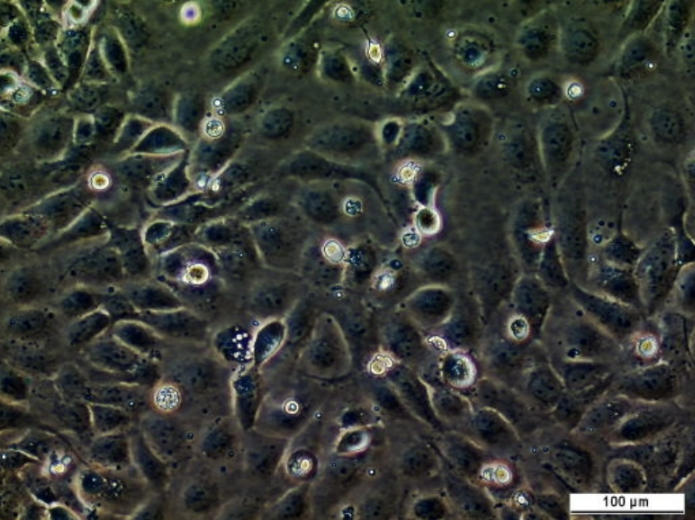
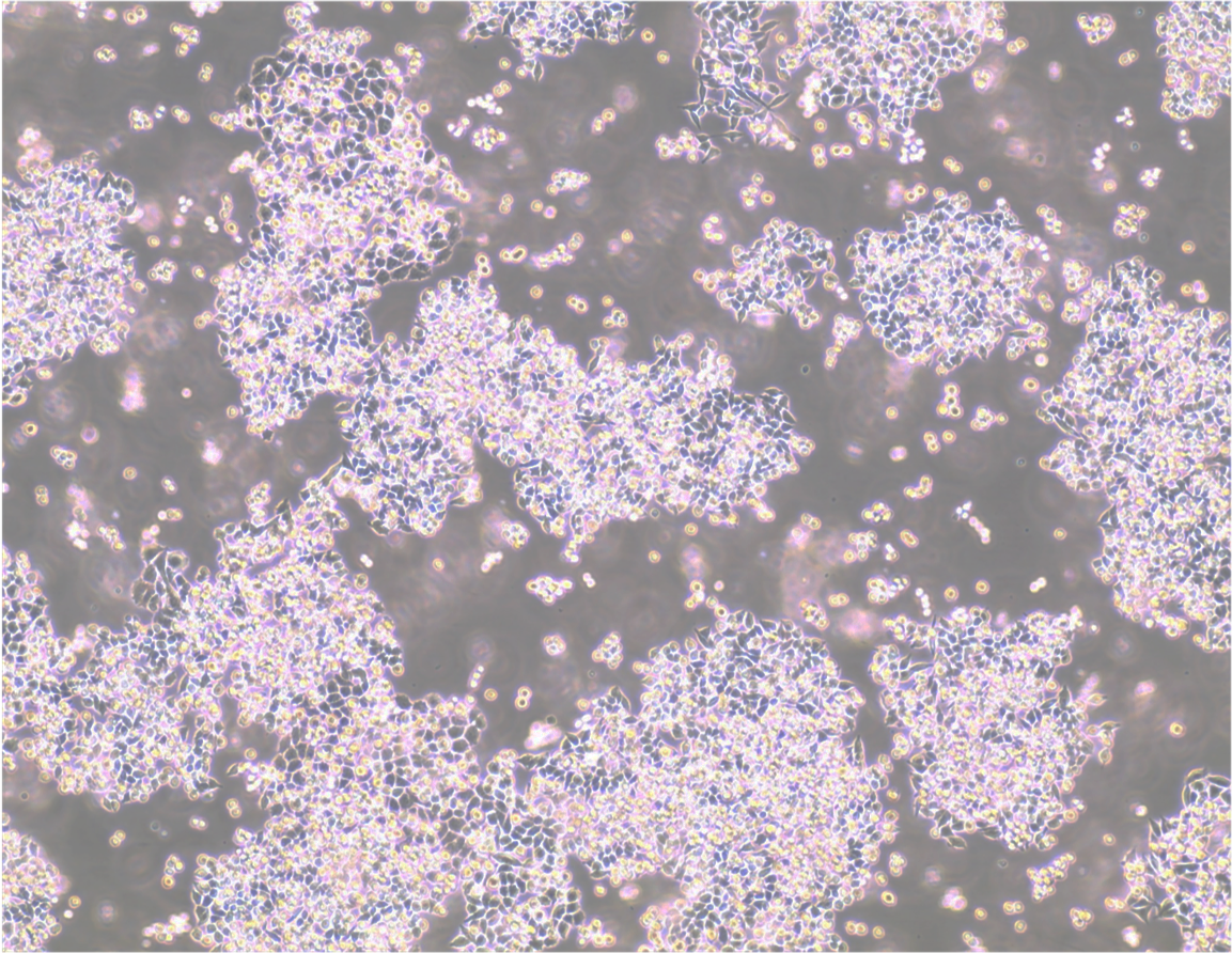
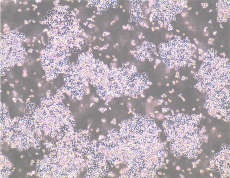
| Morphology: | A spindle-like epithelial morphology, or rounded |
| Growth properties: | The cells do not form a 100% confluent monolayer but at 70% confluence tend to detach in small clumps that float in the media. About 10-20% of the cells will be attached with a spindle-like epithelial morphology, the remainder will appear rounded |
| Primary citation: | Summerhayes et al. 1979. J Natl Cancer Inst. 62(4):1017-23. PMID: 107359 |
| Alternate name: | MB49, MB-49, MB49 Mouse Bladder Carcinoma Cell Line |
|---|---|
| Product description: | The MB49 tumour model is a urothelial carcinoma cell line derived from an adult C57BL/ICRF-a(t) mouse bladder epithelium transformed by chemical carcinogen in culture. It is one of the most-established tumour cell line from mouse (mus musculus) to study human bladder cancer, widely used by scientists for more than 45 years after its original publication by Cancer Research UK researcher – Leonard Franks. MB49 cell line can be used both as an in vitro and in vivo murine model for bladder cancer, thanks to its clinically relevant metastatic potential. MB49 cells also shares pivotal tumour characteristics with human bladder cancer. MB49 cell line key features: (1) Rapidly generates tumours with subcutaneous or orthotopic injections into syngeneic mice (Summerhayes et al. 1979; Kasman et al. 2013). (2) Recapitulates key features of sex differences in bladder tumour growth (White-Gilbertson S, et al. 2016). (3) Loss of the Y-chromosome and expression of male-specific antigens, a frequent feature observed in human bladder cancer (Fabris et al. 2012). (4) Dose-dependent enhanced proliferation to dihydrotestosterone and lack of proliferation to human chorionic gonadotrophin (White-Gilbertson S, et al. 2016). (5) Responsiveness to immune checkpoint inhibitors (Vandeveer et al. 2016) and other agents undergoing clinical investigation (Shingo et al. 2022), making it ideal for testing novel treatment combinations. (6) Model to explore immunogene therapy, such as adenoviral vectors (Loskog et al. 2005). Further accelerate bladder cancer research, with our luminescent derivative of MB49, MB49-luc Cell Line (Cat. #: 161579). The bioluminescence can be detected by in vivo imaging and offers a readout for tumour take, growth and reduction. |
| Gender: | Male |
| CRISPR: | No |
| Conditional: | No |
| Production details: | Derived from adult C57BL/Icrf-a’ mouse bladder epithelial cells via single 24 h treatment with a chemical carcinogen on the second day of primary culture |
| Cellosaurus ID: | CVCL_7076 |
| Model description: | The cells transplanted into syngeneic mice were shown to generate carcinomas |
| Disease: | Cancer;Cancer |
| Format: | Frozen |
|---|---|
| Storage conditions: | Liquid Nitrogen |
| Shipping conditions: | Dry ice |
| Growth medium: | DMEM Complete Medium or in DMEM-High Glucose with 10% FBS and 1X Penicillin/Streptomycin (optional) |
| Subculture routine: | Split sub-confluent cultures (80-85%) 1:6 to 1:10 using Accutase or trypsin-EDTA solution. Cells are cultured at 37°C, in a humidified incubator with 5% CO2. |
| Temperature: | 37° C |
| Atmosphere: | 5%CO2 |
| Storage medium: | Cells can be cryopreserved in growth medium with 10% DMSO using a Nalgene slow freeze Mr.Frosty container. |
| Mycoplasma free: | Yes |
| Biosafety level: | 1 |
| References: |
Albertó et al. 2019. Oncol Lett. 17(3):3141-3150. PMID: 30867744. Plote et al. 2019. Oncoimmunology. 8(5):e1577125. PMID: 31069136. Shi et al. 2019. Onco Targets Ther. 12:4403-4413. PMID: 31239709. White-Gilbertson et al. 2016. Bladder (San Franc). 3(1). PMID: 26998503. Kasman et al. 2013. J Vis Exp. (82):50181. PMID: 24326612. Zhu et al. 2013. BMC Urol. 13:57. PMID: 24188098. Fabris et al. 2012. Cancer Genet. 205(4):168-176. PMID: 22559978. Chen et al. 2009. J Urol. 182(6):2932-2937. PMID: 19853870. Loskog et al. 2005. Lab Anim. 39(4):384-393. PMID: 16197705. Brocks et al. 2005. J Urol. 174(3):1115-1118. PMID: 16094076. Günther et al. 1999. Cancer Res. 59(12):2834-2837. PMID: 10383142. Summerhayes et al. 1979. J Natl Cancer Inst. 62(4):1017-1023. PMID: 107359. |
|---|
Images
View Gallery
Product Images
MB49-provenance-and-ownership-v.3
153368-765×500-1.jpg
MB49
light microscope image of MB49 in culture

Citations And Reviews
 Powered by Bioz
 Related Tools for Cell lines
| Cat. # | Tool Name | |||||||||||||||
|---|---|---|---|---|---|---|---|---|---|---|---|---|---|---|---|---|
| 151479 | HCT 116 p300 KO [D10] Cell Line |
Key Info
HCT 116 p300 KO [D10] Cell Line
|
View Tool | |||||||||||||
| 151480 | HEK293A EGFP-LC3 Cell Line |
Key Info
HEK293A EGFP-LC3 Cell Line
|
View Tool | |||||||||||||
| 151488 | B6.1.1 Mouse ES Cell Line |
Key Info
B6.1.1 Mouse ES Cell Line
|
View Tool | |||||||||||||
| 151663 | MEF PKCe KO KI Cell Line |
Key Info
MEF PKCe KO KI Cell Line
|
View Tool | |||||||||||||
| 151448 | CMT 167 Cell Line |
Key Info
CMT 167 Cell Line
|
View Tool | |||||||||||||