New cell lines: murine melanocyte and melanoblast cell lines from Dr. Elena Sviderskaya

St George’s University of London and the Functional Genomics Cell Bank strengthen their partnership with CancerTools.org to accelerate cancer research.

St George’s University of London (SGUL) and CancerTools.org announce new cell lines from Dr. Elena Sviderskaya, which are now readily available via the CancerTools.org website.

CancerTools.org is a global, non-profit cancer-focused biorepository with a 40+ year history of making cancer research tools accessible such as antibodies, cell lines, mouse models and more to cancer researchers worldwide.

Leading cancer biologists at SGUL are supporting CancerTool.org’s mission to accelerate cancer research through research tools. Since December 2017, CancerTools.org has been storing, producing and shipping SGUL’s scientists’ research tools worldwide. To accelerate cancer research and discoveries globally, SGUL and the Functional Genomics Cell Bank are now making 68 murine melanocyte and melanoblast cell lines, invented by Dr. Elena Sviderskaya, available through CancerTools.org.

“As a non-profit, CancerTools.org is dedicated to accelerating cancer breakthroughs, by ensuring cancer scientists have access to the highest quality research tools. We are delighted to be able to make these valuable cell lines available to the global research community and to continue strengthening our relationship with SGUL and the Functional Genomics Cell Bank.”

James Ritchie, Head of External Innovation, CancerTools.org

Dr. Elena Sviderskaya specialises in pigment cell and melanoma research1 and is the Director of the Functional Genomics Cell Bank1 at St George’s, University of London. The Functional Genomics Cell Bank specialises in mouse melanocyte and melanoblast lines carrying a variety of pigmentary mutations, immortal human melanocytes, melanoma cell lines, and stem cells. Many of these lines are now available through Cancer.Tools.org.

“We are delighted that the cell lines held in the Functional Genomics Cell Bank at St George's are now available via CancerTools.org to researchers globally. These lines are important for cancer biologists as non-cancerous controls for the behaviour of melanoma lines, and are also useful in testing the importance of melanogenesis in the progression of melanoma and other skin cancers.”

Dr. Elena Sviderskaya, Director of the Functional Genomics Cell Bank

About the cell lines:

Melanocytes are the cells in mammals that produce pigment (melanin), colouring the hair, skin and irises. They develop from unpigmented precursors, melanoblasts, and are located in the bottom layer of the skin’s epidermis.

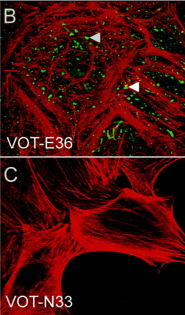
The cell lines deposited with CancerTools.org are immortal melanocyte, melanoblast, and neural-crest stem cell lines derived from embryonic mouse skin. These mutant cell lines are used to study the actions of mutated genes, which affect many body systems besides pigment cells. To date, these cell lines have been used in research on topics including cell differentiation, organelle biosynthesis and transport, protein transport, growth control, cancer and many others.

These cell lines therefore not only add value to many areas of pigment cell research including cell biology, developmental biology, molecular biology, genetics, microscopy, physiology, pathophysiology, ageing and cancer, but also to research involving most major organ systems – eyes, ears, and blood, nervous, respiratory, digestive, excretory and skeletal systems, and disorders such as inflammation, thrombosis and allergy among others.

Colour mutations in mice often have an orthologous mutation in humans with associated pathological effects. There is ready interchange between the advances in pigmentary genetics in the mouse and human, which increases the relevance of these cell lines. Thus, a very broad range of body systems, cellular mechanisms and disorders is addressed by this collection of cell lines.

The majority of cell lines with pigmentary mutations were derived from the C57BL/6J strain mice to exclude confounding differences due to strain background. This is a benefit over human cell lines that have many polymorphisms that can affect biological processes independent of known mutations. Several melanocyte (melan-Ink4a-Arf) lines on the C57BL/6J strain background (genotype a/a) were deposited with CancerTools.org. These and some other deposited lines have mutations at the Ink4a-Arf locus that make spontaneous immortalisation routine. Other lines were derived by rare spontaneous immortalisation. Melan-Ink4a-Arf lines are used in applications like the widely used cell line melan-a that immortalised spontaneously. When mutant cell lines were established from mice of other backgrounds, the corresponding wild-type cell lines were established from littermate controls.

Discover more about Dr. Elena Sviderskaya's cell lines:

Access the cell lines

1https://www.sgul.ac.uk/profiles/elena-sviderskaya#overview

About CancerTools.org

CancerTools.org is the first-of-its-kind non-profit, cancer-focused biorepository where researchers can deposit research tools they have developed in their labs including antibodies, cell lines, organoids, small molecules, mouse models, cell culture media and other state-of- the-art technologies. With our in-house production and global coverage, we can produce, store and supply these tools to fellow scientists in their research to deepen our understanding of cancer and drive innovation.

About St George’s University of London

St George’s, University of London is the UK’s only university dedicated to medical, biomedical and allied health education, training and research. Sharing a clinical environment with a major London teaching hospital in southwest London, our innovative approach to education results in well-rounded and highly skilled clinicians, scientists, and health and social care professionals.

An independent member of the University of London, we have a long and illustrious history of training healthcare professionals, dating back more than 270 years. We are well known for our innovative approach to medical education, being the first UK institution to launch a Graduate Entry Medicine Programme – a four-year fast-track medical degree course open to graduates in any discipline. St Georges’ is the number one university in the UK for Graduate Prospects (on track), according to the Complete University Guide 2024 and second for Graduate Prospects in the recently published Times UK University Rankings for 2024.

Our internationally recognised research delivers cutting-edge scientific discovery through four specialist Research Institutes, directly helping patients through our close links to the clinical frontline and London’s diverse community. We were ranked joint 8th in the country for research impact in the last REF (2021) with 36% of St George’s research assessed as ‘world-leading’ and 100% of our impact cases judged as ‘world-leading’ or ‘internationally excellent.’ Our Institutes focus on biomedical and scientific discovery, advancing the prevention and treatment of disease in the fields of population health, neuroscience, heart disease and infection – four of the greatest challenges to global health in the 21st century.

www.sgul.ac.uk

About the Functional Genomics Cell Bank

The Wellcome Trust has funded a mammalian cell bank (a collection of cell cultures) at St George’s, University of London, in association with the Molecular and Cellular Sciences Section, Neuroscience and Cell Biology Research Institute. The bank specialises in mouse melanocyte and melanoblast lines carrying a variety of pigmentary mutations. Other cell types include immortal human melanocytes, melanoma cell lines, fibroblasts, keratinocytes, mammary epithelial cells, myoblasts, and stem cells.

www.sgul.ac.uk/genomics-cell-bank

Plasmax™ vs DMEM the impact of physiologically relevant cell culture media

Plasmax™

Choosing an appropriate cell culture medium is a crucial step in in vitro cell biology research. With a wide variety of media currently available, finding the correct one for your cell type and particular experiment can be challenging.

Sunada Khadka, a PhD Candidate at MD Anderson Cancer Center, studies cancer metabolism in glioma cells. During her latest research on anaplerosis in glioma cells, Sunada’s initial results obtained in vitro using traditional medium were not reproduced in her in vivo experiments. This led to additional time and resources being used to try and understand the discrepancy.

Here, we explore Sunada’s latest research, and the role PlasmaxTM, a physiologically relevant media, played in resolving the discrepancy between her in vitro and in vivo experimental results.

The researcher

Sunada Khadka

PhD candidate, MD Anderson Cancer Center

Initial in vitro results

Sunada’s research explored the possibility of synergistically killing tumour cells through the inhibition of glycolysis and glutaminolysis, two metabolic pathways that feed The Citric Acid (TCA) cycle.

A novel enolase inhibitor, HEX, was used as a glycolysis inhibitor in this study. HEX was developed through the concept of collateral lethality wherein the passenger deletion of the glycolytic gene ENO1 within a subset of gliomas, selectively renders cancer cells sensitive to inhibition of the redundant isoform ENO2. HEX was tested in combination with CB-839. CB-839 is a glutaminase inhibitor which targets glutamine metabolism and is currently being investigated in randomised clinical trials against a range of malignancies. This made CB-839 of primary interest to extend the metabolism-targeted therapy.

Initially, a pyruvate-free traditional media (DMEM) was used for the in vitro experiments which suggested a very strong effect of CB-839 on ENO1-deleted cancer cells. The combination of CB-839 and HEX provided a dramatic synergetic effect that seemed specific to ENO1-deleted cells.

Plasmax™ bottle

Difficulty recapitulating in vitro results in an in vivo setting

However, when it was attempted to recapitulate the in vitro results in vivo, within an intracranial tumour model, no effect with CB-839 alone and no additive effects with HEX could be seen.

As CB-839 is known to be very poorly permeable across the brain, a subcutaneous in vivo tumour model was used, where Blood Brain Barrier penetration is not an issue. In this case some delay in tumour growth was observed after using CB-839 alone and when used in combination with HEX, but not to the extent seen in the in vitro research.

It was disappointing as we did not see any effect at all after the glutaminase inhibitor and that was very surprising because we saw a very dramatic effect - the complete wipe-out of cells - in vitro.

Sunada Khadka

Figure 1. ENO1-deleted glioma cells (D423) were implanted intracranially in immunocompromised nude mice and tumor growth was monitored weekly across different treatment groups by T2 MRI (indicated by dashed yellow outlines) 20-30 days after tumor implantation. Khadka et al. 2021.

Plasmax™ impact

This inconsistency in data led to a return to in vitro experimental conditions and a closer examination of the cell culture media used. PlasmaxTM was selected as a cell culture media that better reflected the in vivo nutrient profile. PlasmaxTM is a ready-to-use, physiologically relevant cell culture medium, consisting of >80 components, of which >50 have been optimised to levels found within human plasma.

By comparing the  in vitro results from PlasmaxTM to DMEM, it was observed that the toxicity of CB-839 in ENO1-deleted cells is significantly reduced in PlasmaxTM compared to DMEM. These results confirmed the in vivo data and demonstrated that the ENO1-deleted gliomas microenvironment may not be conducive to glutamine addiction.

We decided to try something that matches the physiological profile. And again we saw what we did not expect, which is that the effect of CB-839 seem to be completely diminished in PlasmaxTM medium compared to pyruvate-free DMEM.

Sunada Khadka
ENO1

Figure 2. Sensitivity of glioma cells to CB-839 is attenuated in physiological Plasmax medium. ENO1 homozygously deleted (D423), ENO1-isogenic rescue (D423 ENO1), and ENO1 wild type (LN319) cells were grown in pyruvate free DMEM or Plasmax medium with or without 5 mM pyruvate supplementation. Khadka et al. 2021.

Conclusion:

Sunada’s results emphasize the importance of triaging your cell culture media with physiologically relevant media like PlasmaxTM to better recapitulate the in vivo environment.

As an extension of the paper she recently published, Sunada is now studying the effect of the glycolysis inhibitor in combination with an angiogenesis inhibitor. The restriction of oxygen and nutrient flow into the tumour should have a profound effect. For these experiments she plans on using an intracranial in vivo tumour model and together with PlasmaxTM in her in vitro experiments.

In the future, whatever metabolism related work I do, I'll make sure to compare DMEM to Plasmax™ to ensure that the nutrient profile is not effecting the certain phenotype that I’m seeing. It doesn't hurt - if you are already doing one experiment in one certain media condition, just make another plate with PlasmaxTM for side-by-side comparison. So, I actually don't see why one wouldn’t try it. Especially before you jump into big in vivo experiments, which involve a lot of time and money.

Using physiologically relevant media is a time saver and will make you more confident in your data.

Sunada Khadka

PlasmaxTM is already being repeatedly purchased by various cancer researchers across different academic institutes worldwide. 

Discover how Plasmax™ could benefit your research:

About CancerTools.org

CancerTools.org, the research tools arm of Cancer Research UK, is a non-profit, global community of cancer researchers, academic institutes and societies, with a shared mission to accelerate cancer research discoveries. In this collaborative, researchers contribute research tools and share knowledge to deepen our understanding of cancer, and drive innovation within cancer research.

About Cancer Research UK Glasgow: The Beatson Institute 

One of Cancer Research UK’s core-funded institutes, The Beatson Institute have built an excellent reputation for basic cancer research, including world-class metabolism studies and renowned in vivo modelling of tumour growth and metastasis. Learn more at:  https://www.beatson.gla.ac.uk/About/about-beatson.html

Dr. Wytske M. van Weerden and CancerTools.org

38 new cell lines from Dr. Wytske M. van Weerden now available through CancerTools.org

Image Reference: 2x PTEN -/- mouse prostate cancer cell lines (MuCap system). Histology staining of syngraft tumours (Van Duijn et al., 2018).

Dr. Wytske M. van Weerden is an Associate Professor, who specialises in prostate cancer modelling, with a focus on studying mechanisms of resistance, such as hormone-, chemo- and radio-resistance, with a particular interest in androgen receptor-regulated pathways.1

As part of this research interest, Dr. van Weerden and her team have generated a series of 38 prostate cancer cell lines which are now available through the CancerTools.org collection.

About the cell lines

Prostate cancer is the 2nd most commonly occurring cancer in men and the 4th most common cancer overall. There were more than 1.4 million new cases of prostate cancer in 2020.2 The treatment for prostate cancer focuses on androgen deprivation therapy (ADT) aiming to reduce androgen receptor (AR) activation.3 Although initially effective, in time, resistance develops resulting in castration-resistant prostate cancer (CRPC). Interestingly, the vast majority of clinical CRPC tumours retain a functional AR that continues to drive tumour progression. However, preclinical research of CRPC still relies heavily on AR negative cell line models.4 The research team of Dr. van Weerden has created a comprehensive series of human-derived CRPC cell lines that reflect the changes in AR characteristics very similar to those observed in clinical CRPC. Discover these new cell lines now available through CancerTools.org:

    • PC346C cell line: This cell line has been created from the androgen-responsive xenograft PC346P, a transurethral resection of a primary prostate tumour. This cell line is androgen-responsive and grows slowly in the steroid-stripped medium. It expresses wildtype AR, secretes PSA and is not stimulated by antiandrogen hydroxyflutamide.5


  • PC346C-CRPC panel: The following three cell lines were created by continuously culturing PC346C for over 2 years in various androgen-depleted conditions and show different hormone response properties. The PC346C panel of cell lines is tumorigenic when inoculated subcutaneously in immune-deficient (male) mice.5
  • PC346C-DCC CRPC Cell line: This cell line is unresponsive to both R1881 and hydroxyflutamide and has downregulated AR expression as well as low levels of PSA protein.5
  • PC346C-FLU1 AR overexpressed CRPC Cell line: This cell line grows optimally in steroid-stripped medium or in medium supplemented with hydroxyflutamide, being inhibited by physiologic concentrations of androgens. AR expression is overexpressed in this cell line, which is not a result of AR gene amplification. AR expression is localised in the nucleus. This cell line shows increased AR expression compared to other in vitro 5 The cell secrete PSA.
  • PC346C-FLU2 AR (T877A) mutated CRPC Cell line: This cell line grows optimally in medium supplemented with both R1881 or hydroxyflutamide as it harbours the well-known AR T877A mutation (LNCaP). AR expression in this cell line is localised in the nucleus.5 The cell lines secrete PSA.
    • PC346mAR (T877A) CRPC Cell line: This cell line was established in vitro from the androgen unresponsive PC346I xenograft and shows the AR T877A mutation. This cell line is stimulated by both R1881 and hydroxyflutamide. AR expression in this cell line is localised in the nucleus.5 The cells secrete PSA.
    • 28x PC346C CRPC cell lines: A series of 28 CRPC sublines derived from culturing the source cell line PC346C long-term and continuously under different androgen depleted conditions.4 (see below).

Continue Reading

Six new cell lines from Prof. Michelle Lockley

Prof. Michelle Lockley from Queen Mary University of London, deposits 6 new cell lines with CancerTools.org

Epithelial ovarian cancer is a heterogeneous disease with five different pathological subtypes, the most common being High Grade Serous Cancer (HGSC). Standard treatment to date for epithelial ovarian cancer has been a combination of surgery and platinum-based chemotherapy. However, approximately 80% of patients relapse and respond less well to successive platinum-containing chemotherapy regimens. Platinum-resistance is defined clinically when relapse occurs within 6months of the most recent platinum treatment. Maintenance treatment with PARP inhibitors dramatically improves progression-free survival in platinum-sensitive disease, but new treatments, including PARP inhibitors, have failed to improve survival in platinum-resistant HGSC.

Prof. Michelle Lockley is a Clinician Scientist based at Barts Cancer Institute, Queen Mary University of London. As a consultant medical oncologist specialising in the systemic treatment of gynaecological cancers, she has particular expertise in ovarian cancer.

Pre-clinical research relies on cell-based disease models, but creating permanent cell lines from human HGSC has proven to be challenging and the extent to which these lines reflect characteristic features of relapsed, human HGSC is largely unknown. As part of her research into epithelial ovarian cancer, Michelle and her team used the OVCAR4, Cov318 and Ovsaho cell lines to generate a unique panel of platinum-resistant, in vitro and in vivo, High-grade serous carcinoma (HGSC) models, that recapitulate the genetic and clinical features of human epithelial ovarian cancer.

To make these cell lines available globally to the wider ovarian cancer research scientific community via CancerTools.org, Michelle deposited the following cell lines:

  1. IVRO1 Cell Line
  2. OvsahoCarbo Cell Line
  3. OV4Cis Cell Line
  4. OV4Carbo Cell Line
  5. CovCis Cell Line
  6. OvsahoCis Cell Line

These cell lines are platinum-resistant HGSC models which:

  • share multiple transcriptomic features with relapsed human HGSC
  • have evolved diverse in vivo phenotypes reflecting the human disease
  • share genetic and transcriptional profiles with platinum-resistant human HGSC
  • accurately reproduce the phenotypic diversity seen in patients.

In addition, the infiltrative and metastatic intraperitoneal phenotype produced by Ov4Carbo cells is analogous to the most usual pattern of recurrent, human HGSC.

Discover more about these cell lines:

GATA3-eGFP reporter cell line: Advancing cancer drug discovery and Innovative tools – Insights from Prof. Matthew Holley

The research tool: GATA3-eGFP reporter cell line

GATA3, zinc finger transcription factor, is associated with numerous types of cancer in which its level of expression is critical. Drugs that modulate GATA3 expression are of particular interest, which is why a high-throughput  screening tool is an important addition to the research tool portfolio. Matthew Holley, Emeritus Professor at the University of Sheffield, has shared insights about such one-of-a-kind tool – the GATA3-eGFP reporter cell line, and other in vitro models  he has developed throughout his research- which he has generously contributed to CancerTools.org.

There are no other cell lines with this potential for the study of GATA3 in cancer research.

Emeritus Prof. Holley

The contributor

Emeritus Professor Matthew Holley

University of Sheffield; School of Biosciences

GATA3 in development and cancer

GATA3 is one of the GATA family transcriptional factors; zinc finger proteins that bind the consensus DNA sequence (T/A)GATA(A/G) of a target gene promoter. It is highly conserved among vertebrates and expressed in various tissues and cell lineages, such as immune cells, adrenal glands, placenta, kidneys, skin and breast tissue, inner ear, hair follicles, and nervous system.

GATA3 is responsible for regulating transcription during both development and cell differentiation, and its expression level is crucial at embryonic and postnatal stages. The importance is apparent from examples like early embryonic lethality due to GATA3 deletion, HDR syndrome associated with GATA3 haploinsufficiency, and the fact that Gata3 has been implicated in tumorigenesis. Previous studies have shown that it is involved in T-cell neoplasms, antagonizes cancer progression in PTEN-deficient prostates, and represents a useful marker for luminal category tumours in breast cancer.

GATA3-eGFP reporter cell line for drug discovery

In 2009, Prof. Holley’s group published  results from a gene array analysis they conducted using inner ear cell lines [1].  They showed  a clear, previously unknown, functional link between GATA3 and the Akt signalling pathway; increased activity of which is often associated with tumour progression and therapies resistance. The pathway is essential for regulating cell survival and proliferation, and aberrations in its activation are linked to several human cancers, including breast, lung, ovarian, and prostate cancers.

Prof. Holley delved into exploring how to modulate  gata3  expression, recognising its fundamental role in significant pathways associated with cancer progression as well as in the development of various organs and the nervous system.

Image: Inventing institution: University of Sheffield, UK

The GATA3-eGFP cell line was established by the cross of two well-characterised transgenic mouse lines. One mouse line stably expressed the H-2Kb-tsA58 transgene in which a temperature-sensitive variant of the SV40 large T-antigen was expressed under the control of an inducible promoter driven by gamma-interferon. The second, GATA3eGFP BAC-transgenic mouse line expressed eGFP under the control of the GATA3 promoter. The clones were selected according to the characteristics of the original tissue.

The GATA3-eGFP reporter cell line expresses GATA3-eGFP via an enhancer that is expressed in a pattern very closely resembling that of native GATA3 during mouse embryonic development. Fusion of GATA3 enhancer to eGFP is a key feature of this cell line allowing high-throughput screening of drugs that modulate the expression level of gata3.

First high content screening tests with established GATA3 modulators have shown effectiveness at the anticipated concentrations in the GATA3-eGFP cell line. The tool stands out as the only GATA3 reporter line capable of efficiently screening extrinsic factors influencing GATA3 expression in high-throughput systems.

The key objective of this case study is to show the high potential of this tool for those research laboratories interested in understanding GATA3’s role in various processes in cancer and other diseases.

Other in vitro models

For many years, Prof. Holley dedicated his research efforts to understand development and functioning of the inner ear in mammals, with an emphasis on exploring potential therapies for hearing loss.

Mammalian hair cells located in the inner ear are sensory cells that detect sound, gravity, and acceleration. Progressive loss of the hair cells is one of the main causes of deafness. Hearing loss is widespread and mostly irreversible as the mammalian cochlea is unable to regenerate hair cells that are lost, unlike amphibians and birds.

Mammalian auditory research is complicated by the fact that the very small numbers of auditory sensory epithelial cells do not proliferate postnatally or  in vitro,  which makes them experimentally inaccessible. Thus, Prof. Holley and his colleagues developed a number of  in vitro  models for the differentiation of sensory hair cells and sensory nerves.

The established inner ear cell lines are derived from known cell populations during specific times of mouse inner ear development. They have been shown to correlate with gene expression profiles of the location from which they were derived. The lines are also conditionally immortal and can be grown under proliferating or differentiating conditions.

These cell lines were generously contributed to CancerTools.org and used for exploring the possibility of inner ear regeneration through both cell transplantation and the activation of early developmental processes [2, 3].

I believe in open access to all reagents where possible. They are costly to make and their application can advance research as well as return some investment back to the charitable funding agencies that enabled their production.

Emeritus Prof. Holley

Image: Expression of the β4 integrin subunit in VOT-N33 and VOT-E36. (Adopted from Lawoko-Kerali et. al. 2004. Dev Dyn. 231(4):801-14. PMID: 15499550 Fig.2 B, C) 

Conclusion:

At CancerTools.org  we continue to work with  Cancer scientists who can deposit research tools they have developed in their labs, including cell lines, antibodies,  organoids, mouse models, cell culture media, small molecules and other state-of-the-art technologies, into our biorepository etc.

Access Emeritus Prof. Holley's cell lines:

References

1. Milo et.al. 2009. PLoS One. 4(9):e7144. PMID: 19774072.

2. Rivolta & Holley. 2002. J Neurobiol. 53(2):306-318. PMID: 12382283. 

3. Lawoko-Kerali et. al. 2004. Dev Dyn. 231(4):801-814. PMID: 15499550. 

Tumour Immunology – turning cancer’s weapons against itself

Introduction

For over a century, researchers have been focused on understanding how tumours evade the immune response and thrive. This research has identified key interactions between immune cells and cancer cells, leading to the development of many novel oncology treatments and improved prognosis for many patients.

Image: Light micrograph of a section through a lymph node affected by Hodgkin’s lymphoma, a cancer of lymphoma white blood cells. Stock image.

Tumour immunology encompasses the study of interactions between tumours and the immune system, focusing on understanding how tumours can evade immune responses and thrive. The origin of tumour immunology can be attributed to William Coley, who created the Coley toxin – a heat-inactivated streptococcal cocktail, used to treat sarcomas in the 1890s successfully (1). This treatment was created to stimulate what we now understand to be the immune system to fight cancer. The Coley toxin fell out of favour with the discovery of other oncological treatments such as chemotherapy and radiology (1). This divide in approach between the fields of cancer and immunology continued until around the 90s when fundamental discoveries around inhibiting immune checkpoints were made, ultimately leading to a noble prize for Allison and Honjo in 2018 for their work on cancer immunology (2).

Despite significant advances in the field and the successful implementation of therapeutics that target the immune response, the battle within tumour immunology continues with challenges, such as immunotherapy resistance and efficacy. Here we discuss some of the current barriers within tumour immunology, such as immunotherapy efficacy, the tumour microenvironment (TME), as well as potential avenues being explored to overcome this resistance.

A challenging environment: Why is the tumour microenvironment such a formidable opponent to immunotherapies?

The TME is a complex and dense battleground of non-cancerous cells, signal molecules and structural components that surround and interact with the tumour. It includes immune cells such as macrophages and T cells, as well as stromal cells like cancer-associated fibroblasts (3). A major barrier to successful tumour immunology based treatment is the immunosuppressive TME which contains cells, signals and conditions that restrain the anti-tumour response. The TME facilitates immune evasion through an enrichment of regulatory T cells and M2 macrophages, which can inhibit effector immune cells and secrete immunosuppressive cytokines, including, IL-10 and TGF-B (4). Upregulation of checkpoint proteins like PD-L1 on tumour cells is another cancer escape mechanism used in the TME (3). Immunotherapies like immune checkpoint inhibitors, and CAR T cell therapy, aim to reinvigorate anti-tumour responses to overcome immunosuppression in the TME. Additionally, immune filtration can be an issue due to the increased extracellular matrix in the TME, leading to ‘cold’ tumours (3). As the TME’s immune evasion and immunosuppression is such a significant obstacle to immunotherapies, there is a need to elucidate the mechanisms of action in the TME.

Cancer's signalling sabotage: How intracellular signalling affects tumour immunology?

At the heart of cancer’s defence against the immune system and consequently immunotherapies, lies cancer’s ability to commandeer intrinsic cell signalling pathways that regulate immune recognition and response. Tumours hijack intrinsic signalling pathways to evade immune attacks. Oncogenic mutations to key signalling pathways such as PI3K/mTOR, RAS/RAF/MAPK, Wnt/beta-catenin, STAT3 and NF-kB play dual roles in promoting tumour growth while also mediating an immunosuppressive effect (5). Hyperactivation or dysregulation of these pathways can lead to a decrease in antigen presentation, less T cell infiltration, induction of inhibitory receptors, recruitment of immunosuppressive cells and interference with immune metabolism (5). By understanding dysregulated signalling pathways in tumours, it provides researchers with more targets to drug, to restore immune recognition of tumours.

An infiltration mission: How do we turn ‘cold’ tumours ‘hot’?

Immunotherapies take aim at tumours by harnessing the immune system’s power, but many malignancies reside in immune blind spots, escaping detection. ‘Cold’ tumours occupy these blind spots, as these tumours lack typical immune cell infiltration and inflammation. These ‘cold’ tumours also tend to have a lower mutational burden and display fewer tumour specific antigens (6). Because of the lack of tumour antigens, T-cells are not primed and activated and therefore, do not infiltrate into the TME. Checkpoint inhibitors  work by blocking inhibitory signals on T cells, if no T cells are present due to lack of activation, checkpoint inhibitors have no T cells to target. To tackle these ‘cold’ tumours with modern immunotherapies, we need to turn ‘cold’ tumours ‘hot’. Some potential tactics include combination therapies integrating epigenetic modulators like HDAC inhibitors, which can upregulate MHC class I and II expression on tumour cells, making them better targets for T cells (7).  HDAC inhibitors have also been shown to shift tumour-associated macrophages from an M2 immunosuppressive to an M1 inflammatory subtype (8). Traditional oncological treatments, like radiotherapy, can also play a role in facilitating the conversion of cold to hot tumours; for example, after radiotherapy, endothelial cells express molecules like ICAM1 which facilitates the attraction of immune cells (9). Encouraging the infiltration of immune cells into cold tumours, could help tip the balance in favour of the immune system, leading to the eradication of tumours.

What are the promises and pitfalls of immunotherapies?

Immunotherapy emerged as a revolutionary paradigm shift in our approach to treating cancer, promising to harness the power of the immune system to target and destroy tumours. The development of adoptive cell therapies and checkpoint inhibitors has improved survival for patients with advanced cancers, particularly in NSCLC and melanoma (10). However, the success of immunotherapy has been limited to a subset of patients. Response rates to PD-1 inhibitors can be as much lower depending on the cancer type due to patients failing to respond or developing resistance. Treatment resistance can arise in several ways, such as loss of antigen presentation, induction of T-cell dysfunction and immunosuppressive cell recruitment. Understanding and overcoming resistance to immunotherapy is essential to increase the number of patients who respond to treatment. To rescue the promise of tumour immunotherapies, an interdisciplinary call-to-arms has been enacted. Immunologists and Microbiologists have been unravelling the impact of the microbiome on clinical outcomes, as the gut microbiome regulates the immune system (11). Machine learning has been used to predict the immunogenicity of neoantigens to develop personalised vaccines (12). Clinicians have been combining checkpoint inhibitors with other therapies like HDAC inhibitors, to improve the efficacy of immunotherapy. Gaining a better understanding of resistance mechanisms in unresponsive patients, is critical to rationally designing new approaches to immunotherapy to defeat cancer.

Conclusion

The struggle between the immune system and cancer rages on, shaping the balance between tumour progression and destruction. To better understand and elucidate these interactions, we present a curated collection of antibodies directed at key topics in tumour immunology.

  • Antibodies for immunophenotyping, immune checkpoints, the TME, inflammation, epigenetic changes, and tumour intrinsic signalling pathways.
  • Antibodies directed at a range of innate and adaptive immune cells to assist in immunophenotyping cells.
  • Antibodies directed at both key co-inhibitory and co-stimulatory molecules in immune checkpoint control to study immune checkpoints.
  • Antibodies directed at matrix metalloproteinases, chemokines and hypoxia factors to help understand the interactions in the TME
  • Antibodies directed at epigenetic changes, and key molecules involved in signalling pathways like MYC and p53.

By supplying these tools, we hope to equip scientists with the antibodies they need to gain deeper insights into how cancer evades and suppresses the immune system, and potentially uncover therapeutic targets.

Explore a selection of our immune tumour immunology antibodies:

References

  1. McCarthy et al. 2006. Iowa Orthop J. 26:154-8. PMID: 16789469   
  2. Guo. 2018. BMC Cancer. 18(1):1086. PMID:30415640   
  3. De Visser et al. 2023. Cancer Cell. 13;41(3):374-403. PMID: 36917948 
  4. Tie et al. 2022. J Hematol Oncol. 15(1):61. PMID: 35585567 
  5. Yang et al. 2019. J Hematol Oncol. 27;12(1):125. PMID:31775797  
  6. Wang et al. 2020. MedComm. 26;4(5):e343. PMID: 37638340 
  7. Wang et al. 2020 Protein Cell. 11(7):472-482. PMID: 32162275    
  8. Li et al. Oncogene 2021. 40(10):1836-1850. PMID: 33564072    
  9. Liu et al. 2021. Theranostics. 11,11 5365-5386. PMID: 33859752 
  10. Tiwari et al. 2022. J Biomed Sci. 29, 83. PMID: 36253762 
  11.  Zhang et al. 2023. Exp Hematol Oncol. 28;12(1):84. PMID: 37770953 
  12. Xie et al. 2023. Signal Transduct Target Ther. 6;8(1):9. PMID: 36604431 

Targeting the difficult-to-drug protein – MYC

The research tool: Anti-Omomyc

Cancer is associated with the abnormal growth of cells which proliferate uncontrollably and can metastasize into surrounding tissues. There are over 200 types of cancer and distinct subtypes have been identified. Adding to the challenging complexities of cancer, is tumour heterogeneity, which refers to the differences among tumours of the same type within different patients, among cells within a single tumour, or between a primary and secondary tumour (1). This variability often reduces the efficacy of existing therapies, compromising patient outcomes.

The biotechnological breakthroughs of recent years pushed clinical trials and cancer treatment paradigm to progressively shift from tumour-type centred to precision cancer medicine (2), where the focus is on patient-specific therapies, and challenges like tumour heterogeneity can be overcome through the development of specialized treatments. In contrast, discoveries at the Vall d’Hebron Institute of Oncology (VHIO) support the possibility of a more universal approach, with a single therapeutic option potentially targeting different types of cancer. By taking a universal approach to cancer therapy and targeting the common drivers of cancers across cancer types, this could result in therapies that are universal to cancer patients, but also specific to cancerous cells. The difficulty with this approach, is that these universal targets are often harder to drug, either due to the location or interactions of the target, or because of the universal target’s role in normal cellular function.

CancerTools.org spoke to Dr. Laura Soucek, an investigator at the VHIO and ICREA-born spin-off Peptomyc S.L., and leader of VHIO’s Models of Cancer Therapies Group to find out more.

The contributor

Dr. Laura Soucek

VHIO, ICREA-born spin-off Peptomyc S.L., and leader of VHIO’s Models of Cancer Therapies Group

Targeting a protein altered in many cancer types

Dr. Soucek entered research with one goal in mind – to make a difference in cancer research. To that end, she decided to focus on targeting a protein that was, and still is, considered by many to be “difficult-to-drug” – MYC. The MYC protein sits within the nuclei and acts as a ‘master regulator’ of a variety of cellular functions, including proliferation, differentiation, metabolism, and survival. Due to its multidimensional role, it is critical for MYC to be tightly regulated. Deregulation of MYC leads to altered cellular proliferation and growth, protein synthesis, and metabolism. Additionally, MYC promotes tumour progression through activation of angiogenesis and suppression of the host immune system. However, MYC is a difficult target for cancer therapy. Its location within the nuclei, together with its intrinsically disordered structure and lack of a specific active site, make direct MYC inhibition with traditional strategies challenging. Moreover, all three MYC family members (MYC, MYCN, and MYC) need to be targeted to obtain the most efficient therapeutic impact.

A long road to success

For 20 years, Dr. Soucek has been researching ways to inhibit MYC. In 1998, she designed the dominant-negative form of MYC called Omomyc, as a laboratory tool to study MYC biology. Since then, many milestones have been achieved; first using in vitro systems, transgene expression of Omomyc inhibited MYC activity and reduced proliferation in normal and cancer cells (3). Later, in various animal models of cancer, MYC inhibition by Omomyc exerted remarkable anti-cancer properties, without adverse and irreversible effects (4). Following Omomyc’s successful characterisation as a MYC inhibitor, it is now undergoing clinical development.

When we turned off MYC in cancer cells using Omomyc, we saw that it had a dramatic therapeutic effect in different types of experimental cancer models. The beauty of it is that, while everybody expected normal proliferating cells to suffer too, they simply slowed down their proliferation, but nothing major happened to them. So, we finally had a tool against cancer that seemed not to cause any severe side effects in normal proliferating tissues. And the other thing that really made me happy was that it appeared to be the opposite of personalised medicine: We had a technology that would be applicable to all types of cancers, so that maybe we didn't need different drugs for each of them, maybe we could use a common one for all cancers and patients.

Dr. Soucek
Image: A549 cells with Doxycycline-inducible Omomyc were orthotopically implanted into recipient mice. After treatment with Doxycycline, lung tissue was collected and FFPE lung sections were stained with anti-Omomyc monoclonal [21-1-3].

Omomyc impact

After the promising results obtained in vitro and in vivo, the next step was the conversion of Omomyc to an administrable drug. Dr. Soucek and her team showed that, as an alternative to its use as a transgene, Omomyc could be produced as a recombinant mini-protein. This purified Omomyc mini-protein then showed in Dr.Soucek’s studies that it spontaneously penetrates cancer cells and effectively interferes with MYC transcriptional activity, both in vitro and in vivo (5).

The first Omomyc-derived compound, OMO-103, successfully completed a phase 1 clinical trial in 2022. Previous results, presented at the 34th EORTC-NCI-AACR Symposium, Barcelona, 26-28 October 2022, showed that this first-in-class MYC inhibitor had few side effects, was tolerable, and stabilized disease in some patients. Patients in the trials had a range of solid tumours including pancreatic, bowel, and non-small cell lung cancers, and had received at least three prior lines of therapy (6).

Lasting structural integrity of therapeutic proteins in the target tissue is crucial to maintain their proper function. Since the in vivo stability of these agents can be affected by proteolytic degradation, the validity of Omomyc as the first direct inhibitor of MYC has frequently been questioned (8). A recently published study demonstrated, for the first time, that Omomyc behaves as a stable protein in tumour tissue, with longer lasting structural integrity compared to in blood (7).

Our findings are especially relevant now that Omomyc is being evaluated in clinical trials (Ph1/2 trial) since pharmacokinetic data are usually collected by analysis of blood samples in clinical practice,

Dr. Soucek

Conclusion:

A study published in the journal Genes & Development showed that the expression of Omomyc in preclinical melanoma models disrupts MYC activity and alters gene expression profiles, reducing cancer proliferation and progression (10). This suggests that MYC-targeted therapy by Omomyc could potentially open up a new treatment avenue for melanoma and points to the future development of clinical trials to assess the efficacy of this mini-protein against this tumour type (8). Excitingly, Dr. Laura Soucek’s group has also demonstrated the antimetastatic capacity of Omomyc for the first time in breast cancer (11).

In addition to the Omomyc mini-protein, Dr. Soucek’s laboratory also developed a monoclonal antibody targeting Omomyc, which can be used to study Omomyc levels, as well as better understand its function and effect on biological processes of interest, including cancer.

Omomyc: promising results seen in clinical trials

Discover Dr. Soucek's anti-Omomyc:

Introducing Glucose-free Plasmax™

To further support cell growth in a natural and physiologically relevant environment, we at CancerTools.org are introducing a glucose-free version of Plasmax to complement the original Plasmax formulation.

This defined medium is unique in offering a glucose-free formulation that opens up possibilities for tailored experimental conditions in cancer and cell biology research. It can support a variety of applications including glucose starvation experiments, tumour microenvironment and cancer metabolism studies, metabolic tracing of glucose incorporating stable isotopes (e.g. 13C), which could shed light on critical pathways and mechanisms underlying cancer cell metabolism and glucose utilisation.

While this glucose-free PlasmaxTM eliminates glucose from its formulation, it still preserves the original optimised composition of PlasmaxTM derived from over 80 components. These constituents encompass amino acids and derivatives, inorganic salts, trace elements, and vitamins, with more than 50 components present at levels mirroring those found in human plasma. Hence, the formulation ensures cultured cells closely mimic physiological and metabolic profiles of their in vivo counterparts.

Proven to provide tailored experimental conditions in a physiologically relevant environment, glucose-free PlasmaxTM can support research projects aimed at advancing cancer research and associated metabolic studies.

About CancerTools.org / Cancer Research UK

CancerTools.org, the research tools arm of CRUK, is a non-profit, global community of cancer researchers, academic institutes and societies, with a shared mission to accelerate cancer research. In this collaborative, researchers contribute research tools and share knowledge to deepen our understanding of cancer, and drive innovation within cancer research.
Cancer Research UK (CRUK) is the world’s leading charity dedicated to beating cancer through research. It invests more than £400 million annually into cancer research through funding schemes, conferences, initiatives, resources, and a UK-wide network of research infrastructure across basic, translational, clinical and population research.

Investigating undruggable targets in cancer research

Introduction

Most cancers contain mutations or amplifications of genes. This leads to the production of abnormal proteins, which are responsible for uncontrolled cell growth and the survival of tumour cells. Some of these cancer-driving proteins have been difficult to target pharmacologically and are therefore often referred to as “undruggable” targets. At CancerTools.org, we support basic research in these “undruggable” targets, by making tools developed in these research labs easily available through a single centralised online location.

Read more about difficult-to-drug targets in cancer and explore our selection of relevant reagents developed by key contributors in the field.

What are undruggable targets and why are they relevant to cancer?

Despite numerous tumourigenesis drivers, especially kinases, being successfully used for drug intervention, estimates suggest that 85% of the human proteome is currently undruggable (1). In addition, out of the 5,000 proteins with bindable pockets in the ChEMBL database, only approximately 700 proteins are therapeutic targets for FDA approved drugs (2).

Nevertheless, the notion of undruggable targets has considerably changed over the last few decades and the term is becoming increasingly inaccurate. A few so-called undruggable targets, such as Bcl-2 and RAS, have been successfully targeted with clinically approved inhibitors. Therefore, “difficult to drug” or “yet to be drugged” are more appropriate terms to describe such targets (3).

Intractable targets continue being a key challenge in targeted therapeutics for cancers, together with understanding tumour heterogeneity and resistance mechanisms. Key proteins belonging to difficult-to-drug targets in cancer are often known oncogenes or tumour suppressors. These include transcription factors such as MYC, MYB, p53, STAT3, nuclear factor-κB (NF-κB), and fusion transcription factors frequently associated with paediatric cancers. The RAS family also belong to these proteins, with RAS being mutated in up to 30% of human cancers (4).

Challenges of developing drugs against intractable targets

So far, therapeutic approaches have mostly focused on small molecules and biologics, which target proteins with hydrophobic pockets, those located on the cell surface and those that have been secreted. Due to this approach most potential protein targets, such as transcription factors have been left poorly explored (3,5).

One major challenge with difficult-to-drug proteins arises from the fact that many of them function through protein-protein interactions (PPI). They are often intrinsically disordered proteins, thus relying on their interaction with functional partners and lack a sufficient interaction surface for effective and specific drug binding. In addition to their intracellular localization, these targets frequently lack suitable binding sites that allow inhibition of their functions and play crucial roles in cellular processes such as proliferation and development (3,6,7).

These features have made the drug development for these targets difficult to attain, especially when using conventional therapeutic approaches. However, these challenges have also pushed research boundaries, and the “undruggable” targets of yesterday now represent a promising new area of research.

How researchers are tackling these challenges

Several factors underpin the erosion of the “undruggability” concept, including the development of therapeutics with novel mechanisms of action. These not only include PPI modulators, but also cell therapies, T-cell receptor mimetics and multifunctional small molecules (e.g.  proteolysis-targeting chimeras [PROTACs], peptides/peptidomimetics, nucleic acid–based therapeutics)(7,8,9)*.

A few decades ago, PPIs were commonly regarded as “undruggable”. However, the number of inhibitors approved or under clinical investigation has significantly grown over the years, as the basic knowledge of PPI structure and energetics has expanded. The approval of the Bcl-2 inhibitor, Venclexta, in 2016 for the treatment of chronic lymphocytic leukaemia represented a milestone in this space (10).

The accelerated approval granted by the FDA in 2021, to the first KRAS inhibitor, Sotorasib, (for non-small cell lung cancer with G12C mutation of KRAS) represented a breakthrough in the field, given the previous absence of treatments counteracting the cancer-promoting actions of mutated KRAS proteins (11).

Difficult-to-drug targets, such as MYC, NF-κB and RAS, are essential nodes for cancer maintenance and progression. Pathways regulated by these proteins are often activated in resistant tumours to circumvent the action of targeted therapies, such as kinase inhibitor, and can cause such resistance. Therefore, developing inhibitors that target such proteins is essential to achieve successful inhibition of tumour growth, especially in cancers where an effective targeted therapy is not yet available. Since these nodes serve as a conduit for multiple oncogenic signals, their targeting could also be efficacious in multiple types of cancer, thus reducing the number of agents required (3). 

Importantly, the transition from “undruggable” to “druggable” targets is driven by the availability of structural insights and improved understanding of the biochemical and biological properties of these drug targets. Therefore, continuing basic research in difficult-to-drug targets remains crucial to generate insights and greater understanding into the biological mechanisms of difficult-to-drug targets that can be used in the development of new therapeutic options for cancer patients.

How CancerTools.org is supporting research in difficult-to-drug targets

CancerTools.org supports basic research in difficult-to-drug targets by offerings a selection of relevant tools through a single centralised online location. These include established and novel research tools to investigate MYC, p53, RAS, and Bcl-2. The collection features p53 antibodies created in the laboratory of Prof. Sir David Lane, who discovered this crucial tumour suppressor gene, as well as anti-Omomyc, an antibody developed by Laura Soucek. While the dominant negative Omomyc was first designed as a laboratory tool to study Myc perturbation, it was successively characterised as Myc inhibitor and is currently undergoing clinical development.

Conclusion

Having the right tools to investigate difficult-to-drug targets is crucial to drive technological advancement within the space and has the potential to support a new class of highly targeted cancer treatments, alongside as well as reducing the list of undruggable proteins.

Explore our selection of research tools for difficult-to-drug proteins by target of interest:

Tool enquiry

Please ensure you use your organisation email address rather than personal where possible, as this helps us locate your organisation in our system faster.

Please note we may take up to three days to respond to your enquiry.

CancerTools.org uses the contact information provided to respond to you about our research tools and service. For more information please review our privacy policy.