Category

Antibodies

Tumour Immunology – turning cancer’s weapons against itself

Introduction

For over a century, researchers have been focused on understanding how tumours evade the immune response and thrive. This research has identified key interactions between immune cells and cancer cells, leading to the development of many novel oncology treatments and improved prognosis for many patients.

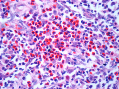
Image: Light micrograph of a section through a lymph node affected by Hodgkin’s lymphoma, a cancer of lymphoma white blood cells. Stock image.

Tumour immunology encompasses the study of interactions between tumours and the immune system, focusing on understanding how tumours can evade immune responses and thrive. The origin of tumour immunology can be attributed to William Coley, who created the Coley toxin – a heat-inactivated streptococcal cocktail, used to treat sarcomas in the 1890s successfully (1). This treatment was created to stimulate what we now understand to be the immune system to fight cancer. The Coley toxin fell out of favour with the discovery of other oncological treatments such as chemotherapy and radiology (1). This divide in approach between the fields of cancer and immunology continued until around the 90s when fundamental discoveries around inhibiting immune checkpoints were made, ultimately leading to a noble prize for Allison and Honjo in 2018 for their work on cancer immunology (2).

Despite significant advances in the field and the successful implementation of therapeutics that target the immune response, the battle within tumour immunology continues with challenges, such as immunotherapy resistance and efficacy. Here we discuss some of the current barriers within tumour immunology, such as immunotherapy efficacy, the tumour microenvironment (TME), as well as potential avenues being explored to overcome this resistance.

A challenging environment: Why is the tumour microenvironment such a formidable opponent to immunotherapies?

The TME is a complex and dense battleground of non-cancerous cells, signal molecules and structural components that surround and interact with the tumour. It includes immune cells such as macrophages and T cells, as well as stromal cells like cancer-associated fibroblasts (3). A major barrier to successful tumour immunology based treatment is the immunosuppressive TME which contains cells, signals and conditions that restrain the anti-tumour response. The TME facilitates immune evasion through an enrichment of regulatory T cells and M2 macrophages, which can inhibit effector immune cells and secrete immunosuppressive cytokines, including, IL-10 and TGF-B (4). Upregulation of checkpoint proteins like PD-L1 on tumour cells is another cancer escape mechanism used in the TME (3). Immunotherapies like immune checkpoint inhibitors, and CAR T cell therapy, aim to reinvigorate anti-tumour responses to overcome immunosuppression in the TME. Additionally, immune filtration can be an issue due to the increased extracellular matrix in the TME, leading to ‘cold’ tumours (3). As the TME’s immune evasion and immunosuppression is such a significant obstacle to immunotherapies, there is a need to elucidate the mechanisms of action in the TME.

Cancer's signalling sabotage: How intracellular signalling affects tumour immunology?

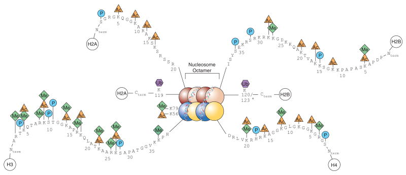
At the heart of cancer’s defence against the immune system and consequently immunotherapies, lies cancer’s ability to commandeer intrinsic cell signalling pathways that regulate immune recognition and response. Tumours hijack intrinsic signalling pathways to evade immune attacks. Oncogenic mutations to key signalling pathways such as PI3K/mTOR, RAS/RAF/MAPK, Wnt/beta-catenin, STAT3 and NF-kB play dual roles in promoting tumour growth while also mediating an immunosuppressive effect (5). Hyperactivation or dysregulation of these pathways can lead to a decrease in antigen presentation, less T cell infiltration, induction of inhibitory receptors, recruitment of immunosuppressive cells and interference with immune metabolism (5). By understanding dysregulated signalling pathways in tumours, it provides researchers with more targets to drug, to restore immune recognition of tumours.

An infiltration mission: How do we turn ‘cold’ tumours ‘hot’?

Immunotherapies take aim at tumours by harnessing the immune system’s power, but many malignancies reside in immune blind spots, escaping detection. ‘Cold’ tumours occupy these blind spots, as these tumours lack typical immune cell infiltration and inflammation. These ‘cold’ tumours also tend to have a lower mutational burden and display fewer tumour specific antigens (6). Because of the lack of tumour antigens, T-cells are not primed and activated and therefore, do not infiltrate into the TME. Checkpoint inhibitors  work by blocking inhibitory signals on T cells, if no T cells are present due to lack of activation, checkpoint inhibitors have no T cells to target. To tackle these ‘cold’ tumours with modern immunotherapies, we need to turn ‘cold’ tumours ‘hot’. Some potential tactics include combination therapies integrating epigenetic modulators like HDAC inhibitors, which can upregulate MHC class I and II expression on tumour cells, making them better targets for T cells (7).  HDAC inhibitors have also been shown to shift tumour-associated macrophages from an M2 immunosuppressive to an M1 inflammatory subtype (8). Traditional oncological treatments, like radiotherapy, can also play a role in facilitating the conversion of cold to hot tumours; for example, after radiotherapy, endothelial cells express molecules like ICAM1 which facilitates the attraction of immune cells (9). Encouraging the infiltration of immune cells into cold tumours, could help tip the balance in favour of the immune system, leading to the eradication of tumours.

What are the promises and pitfalls of immunotherapies?

Immunotherapy emerged as a revolutionary paradigm shift in our approach to treating cancer, promising to harness the power of the immune system to target and destroy tumours. The development of adoptive cell therapies and checkpoint inhibitors has improved survival for patients with advanced cancers, particularly in NSCLC and melanoma (10). However, the success of immunotherapy has been limited to a subset of patients. Response rates to PD-1 inhibitors can be as much lower depending on the cancer type due to patients failing to respond or developing resistance. Treatment resistance can arise in several ways, such as loss of antigen presentation, induction of T-cell dysfunction and immunosuppressive cell recruitment. Understanding and overcoming resistance to immunotherapy is essential to increase the number of patients who respond to treatment. To rescue the promise of tumour immunotherapies, an interdisciplinary call-to-arms has been enacted. Immunologists and Microbiologists have been unravelling the impact of the microbiome on clinical outcomes, as the gut microbiome regulates the immune system (11). Machine learning has been used to predict the immunogenicity of neoantigens to develop personalised vaccines (12). Clinicians have been combining checkpoint inhibitors with other therapies like HDAC inhibitors, to improve the efficacy of immunotherapy. Gaining a better understanding of resistance mechanisms in unresponsive patients, is critical to rationally designing new approaches to immunotherapy to defeat cancer.

Conclusion

The struggle between the immune system and cancer rages on, shaping the balance between tumour progression and destruction. To better understand and elucidate these interactions, we present a curated collection of antibodies directed at key topics in tumour immunology.

  • Antibodies for immunophenotyping, immune checkpoints, the TME, inflammation, epigenetic changes, and tumour intrinsic signalling pathways.
  • Antibodies directed at a range of innate and adaptive immune cells to assist in immunophenotyping cells.
  • Antibodies directed at both key co-inhibitory and co-stimulatory molecules in immune checkpoint control to study immune checkpoints.
  • Antibodies directed at matrix metalloproteinases, chemokines and hypoxia factors to help understand the interactions in the TME
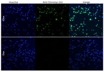
  • Antibodies directed at epigenetic changes, and key molecules involved in signalling pathways like MYC and p53.

By supplying these tools, we hope to equip scientists with the antibodies they need to gain deeper insights into how cancer evades and suppresses the immune system, and potentially uncover therapeutic targets.

Explore a selection of our immune tumour immunology antibodies:

References

  1. McCarthy et al. 2006. Iowa Orthop J. 26:154-8. PMID: 16789469   
  2. Guo. 2018. BMC Cancer. 18(1):1086. PMID:30415640   
  3. De Visser et al. 2023. Cancer Cell. 13;41(3):374-403. PMID: 36917948 
  4. Tie et al. 2022. J Hematol Oncol. 15(1):61. PMID: 35585567 
  5. Yang et al. 2019. J Hematol Oncol. 27;12(1):125. PMID:31775797  
  6. Wang et al. 2020. MedComm. 26;4(5):e343. PMID: 37638340 
  7. Wang et al. 2020 Protein Cell. 11(7):472-482. PMID: 32162275    
  8. Li et al. Oncogene 2021. 40(10):1836-1850. PMID: 33564072    
  9. Liu et al. 2021. Theranostics. 11,11 5365-5386. PMID: 33859752 
  10. Tiwari et al. 2022. J Biomed Sci. 29, 83. PMID: 36253762 
  11.  Zhang et al. 2023. Exp Hematol Oncol. 28;12(1):84. PMID: 37770953 
  12. Xie et al. 2023. Signal Transduct Target Ther. 6;8(1):9. PMID: 36604431 

Targeting the difficult-to-drug protein – MYC

The research tool: Anti-Omomyc

Cancer is associated with the abnormal growth of cells which proliferate uncontrollably and can metastasize into surrounding tissues. There are over 200 types of cancer and distinct subtypes have been identified. Adding to the challenging complexities of cancer, is tumour heterogeneity, which refers to the differences among tumours of the same type within different patients, among cells within a single tumour, or between a primary and secondary tumour (1). This variability often reduces the efficacy of existing therapies, compromising patient outcomes.

The biotechnological breakthroughs of recent years pushed clinical trials and cancer treatment paradigm to progressively shift from tumour-type centred to precision cancer medicine (2), where the focus is on patient-specific therapies, and challenges like tumour heterogeneity can be overcome through the development of specialized treatments. In contrast, discoveries at the Vall d’Hebron Institute of Oncology (VHIO) support the possibility of a more universal approach, with a single therapeutic option potentially targeting different types of cancer. By taking a universal approach to cancer therapy and targeting the common drivers of cancers across cancer types, this could result in therapies that are universal to cancer patients, but also specific to cancerous cells. The difficulty with this approach, is that these universal targets are often harder to drug, either due to the location or interactions of the target, or because of the universal target’s role in normal cellular function.

CancerTools.org spoke to Dr. Laura Soucek, an investigator at the VHIO and ICREA-born spin-off Peptomyc S.L., and leader of VHIO’s Models of Cancer Therapies Group to find out more.

The contributor

Dr. Laura Soucek

VHIO, ICREA-born spin-off Peptomyc S.L., and leader of VHIO’s Models of Cancer Therapies Group

Targeting a protein altered in many cancer types

Dr. Soucek entered research with one goal in mind – to make a difference in cancer research. To that end, she decided to focus on targeting a protein that was, and still is, considered by many to be “difficult-to-drug” – MYC. The MYC protein sits within the nuclei and acts as a ‘master regulator’ of a variety of cellular functions, including proliferation, differentiation, metabolism, and survival. Due to its multidimensional role, it is critical for MYC to be tightly regulated. Deregulation of MYC leads to altered cellular proliferation and growth, protein synthesis, and metabolism. Additionally, MYC promotes tumour progression through activation of angiogenesis and suppression of the host immune system. However, MYC is a difficult target for cancer therapy. Its location within the nuclei, together with its intrinsically disordered structure and lack of a specific active site, make direct MYC inhibition with traditional strategies challenging. Moreover, all three MYC family members (MYC, MYCN, and MYC) need to be targeted to obtain the most efficient therapeutic impact.

A long road to success

For 20 years, Dr. Soucek has been researching ways to inhibit MYC. In 1998, she designed the dominant-negative form of MYC called Omomyc, as a laboratory tool to study MYC biology. Since then, many milestones have been achieved; first using in vitro systems, transgene expression of Omomyc inhibited MYC activity and reduced proliferation in normal and cancer cells (3). Later, in various animal models of cancer, MYC inhibition by Omomyc exerted remarkable anti-cancer properties, without adverse and irreversible effects (4). Following Omomyc’s successful characterisation as a MYC inhibitor, it is now undergoing clinical development.

When we turned off MYC in cancer cells using Omomyc, we saw that it had a dramatic therapeutic effect in different types of experimental cancer models. The beauty of it is that, while everybody expected normal proliferating cells to suffer too, they simply slowed down their proliferation, but nothing major happened to them. So, we finally had a tool against cancer that seemed not to cause any severe side effects in normal proliferating tissues. And the other thing that really made me happy was that it appeared to be the opposite of personalised medicine: We had a technology that would be applicable to all types of cancers, so that maybe we didn't need different drugs for each of them, maybe we could use a common one for all cancers and patients.

Dr. Soucek
Image: A549 cells with Doxycycline-inducible Omomyc were orthotopically implanted into recipient mice. After treatment with Doxycycline, lung tissue was collected and FFPE lung sections were stained with anti-Omomyc monoclonal [21-1-3].

Omomyc impact

After the promising results obtained in vitro and in vivo, the next step was the conversion of Omomyc to an administrable drug. Dr. Soucek and her team showed that, as an alternative to its use as a transgene, Omomyc could be produced as a recombinant mini-protein. This purified Omomyc mini-protein then showed in Dr.Soucek’s studies that it spontaneously penetrates cancer cells and effectively interferes with MYC transcriptional activity, both in vitro and in vivo (5).

The first Omomyc-derived compound, OMO-103, successfully completed a phase 1 clinical trial in 2022. Previous results, presented at the 34th EORTC-NCI-AACR Symposium, Barcelona, 26-28 October 2022, showed that this first-in-class MYC inhibitor had few side effects, was tolerable, and stabilized disease in some patients. Patients in the trials had a range of solid tumours including pancreatic, bowel, and non-small cell lung cancers, and had received at least three prior lines of therapy (6).

Lasting structural integrity of therapeutic proteins in the target tissue is crucial to maintain their proper function. Since the in vivo stability of these agents can be affected by proteolytic degradation, the validity of Omomyc as the first direct inhibitor of MYC has frequently been questioned (8). A recently published study demonstrated, for the first time, that Omomyc behaves as a stable protein in tumour tissue, with longer lasting structural integrity compared to in blood (7).

Our findings are especially relevant now that Omomyc is being evaluated in clinical trials (Ph1/2 trial) since pharmacokinetic data are usually collected by analysis of blood samples in clinical practice,

Dr. Soucek

Conclusion:

A study published in the journal Genes & Development showed that the expression of Omomyc in preclinical melanoma models disrupts MYC activity and alters gene expression profiles, reducing cancer proliferation and progression (10). This suggests that MYC-targeted therapy by Omomyc could potentially open up a new treatment avenue for melanoma and point to the future development of clinical trials to assess the efficacy of this mini-protein against this tumour type (8).

In addition to the Omomyc mini-protein, Dr. Soucek’s laboratory also developed a monoclonal antibody targeting Omomyc, which can be used to study Omomyc levels, as well as better understand its function and effect on biological processes of interest, including cancer.

Omomyc: promising results seen in clinical trials

Discover Dr. Soucek's anti-Omomyc:

Investigating undruggable targets in cancer research

Introduction

Most cancers contain mutations or amplifications of genes. This leads to the production of abnormal proteins, which are responsible for uncontrolled cell growth and the survival of tumour cells. Some of these cancer-driving proteins have been difficult to target pharmacologically and are therefore often referred to as “undruggable” targets. At CancerTools.org, we support basic research in these “undruggable” targets, by making tools developed in these research labs easily available through a single centralised online location.

Read more about difficult-to-drug targets in cancer and explore our selection of relevant reagents developed by key contributors in the field.

What are undruggable targets and why are they relevant to cancer?

Despite numerous tumourigenesis drivers, especially kinases, being successfully used for drug intervention, estimates suggest that 85% of the human proteome is currently undruggable (1). In addition, out of the 5,000 proteins with bindable pockets in the ChEMBL database, only approximately 700 proteins are therapeutic targets for FDA approved drugs (2).

Nevertheless, the notion of undruggable targets has considerably changed over the last few decades and the term is becoming increasingly inaccurate. A few so-called undruggable targets, such as Bcl-2 and RAS, have been successfully targeted with clinically approved inhibitors. Therefore, “difficult to drug” or “yet to be drugged” are more appropriate terms to describe such targets (3).

Intractable targets continue being a key challenge in targeted therapeutics for cancers, together with understanding tumour heterogeneity and resistance mechanisms. Key proteins belonging to difficult-to-drug targets in cancer are often known oncogenes or tumour suppressors. These include transcription factors such as MYC, MYB, p53, STAT3, nuclear factor-κB (NF-κB), and fusion transcription factors frequently associated with paediatric cancers. The RAS family also belong to these proteins, with RAS being mutated in up to 30% of human cancers (4).

Challenges of developing drugs against intractable targets

So far, therapeutic approaches have mostly focused on small molecules and biologics, which target proteins with hydrophobic pockets, those located on the cell surface and those that have been secreted. Due to this approach most potential protein targets, such as transcription factors have been left poorly explored (3,5).

One major challenge with difficult-to-drug proteins arises from the fact that many of them function through protein-protein interactions (PPI). They are often intrinsically disordered proteins, thus relying on their interaction with functional partners and lack a sufficient interaction surface for effective and specific drug binding. In addition to their intracellular localization, these targets frequently lack suitable binding sites that allow inhibition of their functions and play crucial roles in cellular processes such as proliferation and development (3,6,7).

These features have made the drug development for these targets difficult to attain, especially when using conventional therapeutic approaches. However, these challenges have also pushed research boundaries, and the “undruggable” targets of yesterday now represent a promising new area of research.

How researchers are tackling these challenges

Several factors underpin the erosion of the “undruggability” concept, including the development of therapeutics with novel mechanisms of action. These not only include PPI modulators, but also cell therapies, T-cell receptor mimetics and multifunctional small molecules (e.g.  proteolysis-targeting chimeras [PROTACs], peptides/peptidomimetics, nucleic acid–based therapeutics)(7,8,9)*.

A few decades ago, PPIs were commonly regarded as “undruggable”. However, the number of inhibitors approved or under clinical investigation has significantly grown over the years, as the basic knowledge of PPI structure and energetics has expanded. The approval of the Bcl-2 inhibitor, Venclexta, in 2016 for the treatment of chronic lymphocytic leukaemia represented a milestone in this space (10).

The accelerated approval granted by the FDA in 2021, to the first KRAS inhibitor, Sotorasib, (for non-small cell lung cancer with G12C mutation of KRAS) represented a breakthrough in the field, given the previous absence of treatments counteracting the cancer-promoting actions of mutated KRAS proteins (11).

Difficult-to-drug targets, such as MYC, NF-κB and RAS, are essential nodes for cancer maintenance and progression. Pathways regulated by these proteins are often activated in resistant tumours to circumvent the action of targeted therapies, such as kinase inhibitor, and can cause such resistance. Therefore, developing inhibitors that target such proteins is essential to achieve successful inhibition of tumour growth, especially in cancers where an effective targeted therapy is not yet available. Since these nodes serve as a conduit for multiple oncogenic signals, their targeting could also be efficacious in multiple types of cancer, thus reducing the number of agents required (3). 

Importantly, the transition from “undruggable” to “druggable” targets is driven by the availability of structural insights and improved understanding of the biochemical and biological properties of these drug targets. Therefore, continuing basic research in difficult-to-drug targets remains crucial to generate insights and greater understanding into the biological mechanisms of difficult-to-drug targets that can be used in the development of new therapeutic options for cancer patients.

How CancerTools.org is supporting research in difficult-to-drug targets

CancerTools.org supports basic research in difficult-to-drug targets by offerings a selection of relevant tools through a single centralised online location. These include established and novel research tools to investigate MYC, p53, RAS, and Bcl-2. The collection features p53 antibodies created in the laboratory of Prof. Sir David Lane, who discovered this crucial tumour suppressor gene, as well as anti-Omomyc, an antibody developed by Laura Soucek. While the dominant negative Omomyc was first designed as a laboratory tool to study Myc perturbation, it was successively characterised as Myc inhibitor and is currently undergoing clinical development.

Conclusion

Having the right tools to investigate difficult-to-drug targets is crucial to drive technological advancement within the space and has the potential to support a new class of highly targeted cancer treatments, alongside as well as reducing the list of undruggable proteins.

Explore our selection of research tools for difficult-to-drug proteins by target of interest:

Tumour Antigens

Introduction

CancerTools.org is committed to empower cancer researchers and the advancement of novel discoveries in tumour immunology by providing relevant research tools, including antibodies targeting tumour antigens.

Image: Human breast cancer stained with an antibody to Pax-2. Photo credit: Jason Carroll at the CRI.

Tumour antigens are proteins, glycoproteins, glycolipids, or carbohydrates found in tumour cells and recognised by cellular or humoral effectors of the immune system. Tumour antigens are not only useful in identifying cancer cells, they also can be leveraged as targets in cancer therapy. With the ever-increasing potential of tumour antigens in immunotherapy approaches, including cell therapy and cancer vaccines, their discovery and research has become crucial to support successful immunotherapy.

Classification of tumour antigens

Tumour antigens can be classified into tumour-specific antigens and tumour-associated antigens, which in turn can be sub-categorised, based on antigen origin (1,2).

Tumour-specific antigens are restricted to tumours and are not found in healthy cells as they are the result of malignant mutations or the expression of viral elements (1,2). They include:

    >

  • Neoantigens, produced as a direct consequence of genetic alteration caused by tumour DNA mutations and are patient-specific (3)
  • Oncoviral antigens, derived from tumorigenic transforming viruses (4)
  • Endogenous retroviruses (ERVs), fragments of genomic DNA derived from integration of retrotranscribed retroviral RNAs that infected the germ line cells of humans’ ancestors (5)

Tumour-associated antigen have a higher expression level in tumours compared to normal tissue and are shared among several tumours (1,2). They include:

  • Antigens derived from genes overexpressed in tumours, which comprise a class of normal self-proteins minimally expressed by healthy tissues but constitutively overexpressed in cancer cells, e.g., EGFR, hTERT, p53, and carbonic anhydrase IX
  • Differentiation antigens, which are expressed only at specific phases of differentiation of a cell type in healthy tissues and in a specific type of tumour
  • Antigens derived from cancer germline, which are expressed in various tumours but not in normal tissue, except testis and placenta, e.g., cancer testis antigens and oncofoetal antigens

Tumour antigens that are highly expressed by cancer cells and provide information about tumour aggression, metastasis, and treatment responsiveness can also be classified as tumour markers. While tumour markers are not the primary modalities for cancer diagnosis, they can be used as laboratory test to support diagnosis and/or provide information about a specific cancer (6).

Conclusion

Our offering includes antibodies targeting tumour-specific antigens (e.g., oncoviral antigens and neoantigens), tumour-associated antigens (e.g., cancer germline antigens), as well as tumour markers, including those for tumour characterisation and supporting diagnosis.

References

  1. Feola S et al. 2020. Cancers. 12(6): 1660. PMID: 32585818.
  2. Ilyas S et al. 2015. J Immunol. 195(11):5117-22. PMID: 26589749.
  3. Schumacher et al. 2019. Annu Rev Immunol. PMID: 30550719.
  4. Hollingsworth R et al. 2019. NPJ PMID: 30774998.
  5. Bannert N et al. 2018. Front Microbiol. 9:178. PMID: 29487579.
  6. Tumour markers factsheet, National Cancer Institute, NIH

Brainbow Antibodies

The research tool: Brainbow antibodies

Brainbow antibodies were generated by Dawen Cai, PhD, at University of Michigan, to primarily use as part of multicolour labelling strategy for fluorescent imaging of neuronal circuits and individual neurons in mice, Drosophila, and zebrafish and non-neuronal cells in mice.

The contributor

Dr. Dawen Cai

University of Michigan

Multicolour labelling strategy for fluorescent imaging

The Brainbow technology uses stochastic and combinatorial expression of fluorescent proteins to generate colour patterns, which serves as unique identification tag in cells and anatomically complex tissues, such as central nervous system. Dawen Cai, et al. (1) generated a refinement of the Brainbow technology (Brainbow 3) overcoming some of the limitations of the initial approach, such as suboptimal fluorescence intensity, failure to fill all axonal and dendritic processes, and disproportionate expression of the non-recombined fluorescent protein in the transgene. Brainbow 3 includes transgenic murine lines and adeno-associated virus (AAV). This version can also label delicate axonal and dendritic processes by using farnesylated derivatives of fluorescent proteins.

Brainbow applications*

  • Lineage labelling of neurons and non-neuronal cells in mice, e.g., Confetti1 (2) and Ubow (3) models
  • Cell tracing and lineage analysis in zebrafish, e.g., Zebrabow (4) and Drosophila, e.g., dBrainbow (5)
  • Short-term cell labelling in different species via somatic expression, e.g., Brainbow AAV (5)
  • Barcoding of somatic mutations and visualisation of clonal expansion and spread of oncogenes in the Crainbow mouse model (6)
Image: Neurons and interneurons labelled with Brainbow AAV injected into cortex of PV-Cre mice. Antibody amplified mTFP and EYFP are in green, TagBFP is in blue and mCherry is in red. From: Cai et al. 2013. Nat Methods. 10(6):540-7. PMID: 23817127.

Conclusion: Brainbow antibodies

  • Custom-made polyclonal antibodies with specificity for eight different fluorescent proteins
  • Fluorescent proteins derived from organisms of different species (except for EGFP and YFP) to increase specificity and avoid cross-reactivity
  • Antibodies raised against different host species (e.g., chicken, rabbit, rat, and Guinea pig) to allow broader choice of secondary antibodies
  • Antibodies can be used regardless of the sample species, as they react to the relevant fluorescent protein
  • Use is recommended when the endogenous fluorescence of Brainbow is too weak, such as after fixation in histological analysis, to amplify the fluorescent signal

Explore the brainbow antibodies by the fluorescent proteins they target:

References

(1) Cai et al. 2013. Nat Methods. 10(6): 540–547. PMID: 23817127
(2) Snippert et al. 2010. Cell. 143(1):134-44. PMID: 20887898
(3) Ghigo et al. 2013. J Exp Med. 210(9): 1657–1664. PMID: 23940255
(4) Pan et al. 2013. Development. 140(13): 2835–2846. PMID: 23757414
(5) Hampel et al. 2011. Nat methods. 8(3): 253–259. PMID: 21297621
(6) Boone PG, et al. 2019. Nat Commun. 10(1):5490. PMID: 31792216

*List is not exhaustive

Discovering the world of histone modifications and their impact on cancer research

Introduction

Post-translational modifications (PTMs) impact eukaryotic cell diversity by altering protein activity and coordinating cell signalling networks. PTMs are the chemical changes that occur after DNA has been transcribed by RNA into a protein. Defects within PTMs, can cause numerous diseases including:

By increasing our understanding of the mechanisms of PTMs, we can develop new treatments for these diseases.

Image: A diagram showing a full nucleosome with including the histone PTMs on their respective histones. Taken from Keppler, Brian & Archer, Trevor. (2008). Chromatin-modifying enzymes as therapeutic targets – Part 1. Expert opinion on therapeutic targets. 12. 1301-12. 10.1517/14728222.12.10.1301.

PTMs play a crucial role in histone modifications, as they regulate the ability of histones to condense DNA from single strands to tightly wrapped super coiled DNA within nucleosomes. These nucleosomes consist of two dimers of histone H2A-H2B and a tetramer of H3-H4 histones (see Fig.1) and all contain a simple helix turn helix motif and a long amino acid “tail”. PTMs to the long amino acid “tail” change the histone’s function, leading to histone involvement in transcription, DNA repair and replication. These modifications also form the basis of the five different histone PTM families. This article will focus on these five histone PTM families and their associated impacts:

Methylation

Histone methylation is a common PTM that causes differing numbers of methyl groups to attach to a single histone amino acid. Errors in the location and number of methyl groups bonded to this amino acid lead to developmental defects, increased cancer progression and alterations in the DNA repair pathway. Recently Zheng et al., have shown that two methyltransferases (MLL3 and MLL4) associated with mono-methylation of H3K4, have distinct mechanisms, with MLL3 found in gastrulation and MLL4 in lung development. Based on this research, specific inhibitors or activators targeted to each transferase can be designed, providing unique tools to use in precision medicine targeting blood-cell and developmental disorders.

Gene expression is continually suppressed and increased through the activity of MLL methyltransferases, and demethylases. One well characterised demethylase, KDM4, is now a promising target in cancer therapeutics. Disfunction of the KDM4 enzyme leads to sustained proliferation and genomic instability, which in breast cancer can be overexpressed by approximately 60%. This over-expression means KDM4 inhibitors are an interesting therapeutic target with new chemicals being studied regularly for their ability to rebalance gene expression.

Acetylation

Knowledge of histone acetylation and deacetylation has become important in understanding transcriptional regulation. In recent years, inhibitors of histone deacetylases (HDAC), which cause genetic repression through the removal of acetyl groups, have become popular in cancer drug development. For example, in patients with stage 4 neuroblastoma, over-expression of HDAC10 leads to cytotoxic drug resistance resulting in poor survivability of a patient. 

Research has shown that the inhibition of this protein increases cell sensitisation to these cytotoxic drugs, which could result in higher numbers of successful cancer treatmentsCommon essential genes found within this drug resistance pathway, are also found within apoptosis. These genes need to be tightly regulated using acetylation and deacetylation, otherwise uncontrollable cell proliferation occurs. In December 2020, Rajan et al., published a review into Histone Acetylation-/Methylation-Mediated Apoptotic Gene Regulation in Hepatocellular Carcinoma, showing that an increase in  acetylation on histone H2B, H4 and hypoacetylation of histone H4 leads to a progression in cellular apoptosis. This can also be seen when the loss of H4K16 deacetylati

Knowledge of histone acetylation and deacetylation has become important in understanding transcriptional regulation. In recent years, inhibitors of histone deacetylases (HDAC), which cause genetic repression through the removal of acetyl groups, have become popular in cancer drug development. For example, in patients with stage 4 neuroblastoma, over-expression of HDAC10 leads to cytotoxic drug resistance resulting in poor survivability of a patient.

Research has shown that the inhibition of this protein increases cell sensitisation to these cytotoxic drugs, which could result in higher numbers of successful cancer treatments. Common essential genes found within this drug resistance pathway, are also found within apoptosis. These genes need to be tightly regulated using acetylation and deacetylation, otherwise uncontrollable cell proliferation occurs. In December 2020, Rajan et al., published a review into Histone Acetylation-/Methylation-Mediated Apoptotic Gene Regulation in Hepatocellular Carcinoma, showing that an increase in acetylation on histone H2B, H4 and hypoacetylation of histone H4 leads to a progression in cellular apoptosis. This can also be seen when the loss of H4K16 deacetylation specifically causes inhibition and deviation from the normal cell death pathway in Non-alcoholic Steatohepatitis. By increasing our understanding of HDACs and their inhibitors, advancements in cancer treatments can be achieved that build on the already successful therapeutic toolbox.

on specifically causes inhibition and deviation from the normal cell death pathway in Nonalcoholic Steatohepatitis. By increasing our understanding of HDACs and their inhibitors, advancements in cancer treatments can be achieved that build on the already successful therapeutic toolbox.  

Ubiquitylation and Sumoylation

It is estimated that 5–15% of H2A and 1–2% of H2B are conjugated with ubiquitin in vertebrate cells. Ubiquitination plays a role in the regulation of transcription and histone ubiquitylation can define a histone’s activity and protein-protein interactions. In April 2021, cross talk between ubiquitylation, acetylation and methylation was identified. DOT1, a methyltransferase with involvement in leukaemia, had its activity increased by H4K16 acetylation and H2B ubiquitylation. This fundamental research provides further mechanistic detail into leukaemia development that may result in cancer therapeutics.

Histone SUMOylation occurs on all four core histones, the linker histone H1 and histone variants H2A.Z and H2A.X. Although it was discovered over a decade ago, the direct effects of histone SUMOylation are only recently being explored. While histone SUMOylation was originally designated as a marker of transcription repression by competing with other PTMs on histones, it has now been established that SUMOylation is a fine tuner of transcription and DNA damage where it can both act as a repressor or activator. Discussions on druggable sumoylation factories that provide opportunities for clinical interventions within cancers, neurodegeneration and viral infections are now taking place.

Phosphorylation

Histone phosphorylation plays a major role in both transcription and DNA repair. A common measurement of DNA damage is the amount of histone H2A.XS139phospho (γH2AX) present within a cell. γH2AX notifies the cell of a double stranded break and results in downstream signalling of repair proteins and cell cycle stalling. This use of γH2AX as a measuring tool of DNA damage has allowed studies into glomerular diseases where type VI collagen can be deposited as large nodular lesions within the body.

As well as being used as a signal of DNA damage, the actual repair mechanism and signalling pathways linked to γH2AX are still being researched. A recent Nature Communications publication by Dobersch et al., shows that the presence of H2A.X phosphorylation at sites of DNA damage, removes the methylation groups off DNA, allowing for transcriptional activity at the gene of interest. This adds further mechanistic detail to an already complex process of which comes first, PTMs on a histone or the recruitment of proteins associated with sensing DNA damage. By answering this question, researchers can improve the cells ability to fix damaged cells before they become oncogenic.

Conclusion

Whilst PTMs on histones have been heavily characterised, their roles have only become more complex, with some both activating and repressing transcription. With 8 histones making a nucleosome and 30 million nucleosomes per cell, the cross talk between all these PTMs is complex. This article shares only a glimpse into the latest research, highlighting a few of the new applications and potential uses of histone PTMs. The increased focus on histone PTMs and the subsequent discoveries could help provide new avenues into potential treatments for various cancers, and other associated diseases in the near future.

Explore the histone related tools in our collection:

Tool enquiry

Please ensure you use your organisation email address rather than personal where possible, as this helps us locate your organisation in our system faster.

CancerTools.org uses the contact information provided to respond to you about our research tools and service. For more information please review our privacy policy.