Cat. #153422
H357 Cell Line
Cat. #: 153422
Unit size: 1x10^6 cells / vial
Availability: 10-12 weeks
Tissue: Tongue
Disease: Cancer
Model: Tumourigenic cell line
£575.00
This fee is applicable only for non-profit organisations. If you are a for-profit organisation or a researcher working on commercially-sponsored academic research, you will need to contact our licensing team for a commercial use license.
Contributor
Inventor: Stephen Prime
Institute: University of Bristol
Tool Details
*FOR RESEARCH USE ONLY (for other uses, please contact the licensing team)
- Name: H357 Cell Line
- Alternate name: H-357; h357
- Research fields: Cancer
- Tissue: Tongue
- Disease: Cancer
- Growth properties: Adherent
- Model: Tumourigenic cell line
- Conditional: Yes
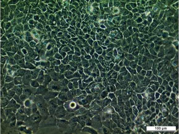
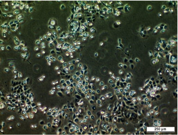
- Description: H357 was established from a squamous cell carcinoma (SCC) of the tongue (20mm) of a 74 year-old male patient. H357 has the following features: polygonal morphology, STNMP stage I, well differentiated and node negative tumour. This cell line has mutant p53, codon 110 exon 4, G to A; the previously reported mutant Ha-ras status of this cell line: codon: 13, G to A, codon 61, A-G is under investigation. H357 is highly responsive to TGF beta and undergoes epithelial to mesenchymal transition. H357 cells are tumourigenic in athymic nude mice on subcutaneous injection and non-tumourigenic on orthotopic injection. Haplotype information: A*02,A*31; B*40,B*44; Cw*03,Cw*05. Explore more human oral SCC cell lines from the same inventor including: H157, H376, H400, H314 and H103.
- Cellosaurus id: CVCL_2462
Target Details
- Target: Human oral squamous cell carcinoma, tongue
Handling
- Format: Frozen
- Growth medium: DMEM:HAMS F12 (1:1) + 2mM Glutamine + 10% Foetal Bovine Serum (FBS) + 0.5 ug/ml sodium hydrocortisone succinate.
- Unit size: 1x10^6 cells / vial
- Shipping conditions: Dry ice
- Subculture routine: Split sub-confluent cultures (70-80%), approximately every 5-6 days, 1:8 to 1:10 using 0.05% trypsin/EDTA; 5% CO₂; 37°C. Suggested seeding density of 5 x 1000 cells/cm². Cells can take approximately 10 minutes to detach, an alternative is to trypsinise 2 to 3 times with fresh trypsin for shorter periods for each trypsin application. Avoid knocking flasks during the trypsinisation process as this can lead to loss of viability.
References
- Fahey et al. 1996. Br J Cancer. 74(7):1074-80. PMID: 8855977.
- Yeudall et al. 1995. Eur J Cancer B Oral Oncol. 31(2):136-143. PMID: 7633286.
- Prime et al. 1994. Int J Cancer. 56(3):406-412. PMID: 7508893.
- Prime et al. 1994. Br J Cancer. 69(1):8-15. PMID: 8286215.
- Yeudall et al. 1993. Eur J Cancer B Oral Oncol. 29B(1):63-67. PMID: 8180579.
- Prime et al. 1990. J Pathol. 160(3):259-269. PMID: 1692339.


