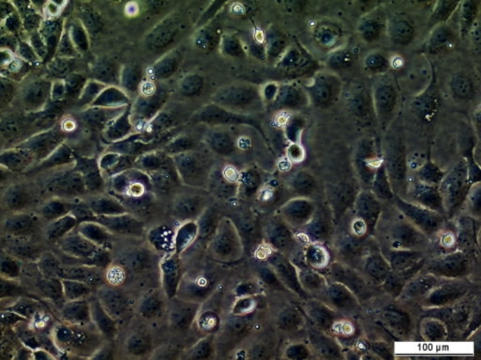
HEK293-NLuc (control) cell line
Endoplasmic reticulum (ER) stress is caused by the accumulation of unfolded proteins in the ER, which leads to the activation of unfolded protein response (UPR) through three transmembrane protein sensors located in the ER membrane. The sensors correspond to three branches of the UPR, namely protein kinase RNA-like endoplasmic reticulum kinase (PERK), activating transcription factor […]
Contributors
| Inventor | Institute |
|---|---|
| Tina Sket, Andrii Domanskyi | University of Helsinki, Institute of Biotechnology, HiLIFE |
| Cat. #: | 161711 |
|---|---|
| Tool sub type: | Continuous |
| Research Fields: | Cell biology |
| Organism: | Human |
| Tissue: | Kidney |
| Model: | Knock-In |
| Morphology: | Epithelial |
| Growth properties: | Adherent |
| Product description: | Endoplasmic reticulum (ER) stress is caused by the accumulation of unfolded proteins in the ER, which leads to the activation of unfolded protein response (UPR) through three transmembrane protein sensors located in the ER membrane. The sensors correspond to three branches of the UPR, namely protein kinase RNA-like endoplasmic reticulum kinase (PERK), activating transcription factor 6 (ATF6), and inositol-requiring enzyme 1 (IRE1) branches. Upon ER stress, IRE1 dimerizes and oligomerizes, and its endonuclease domain is activated. It specifically targets X-box-binding protein 1 (XBP1) mRNA, from which a 26 nt intron is spliced. This allows a complete translation of spliced XBP1 mRNA into a functional protein that acts as a transcription factor. Together with the other pathways, the UPR leads to a decrease in the protein folding load by causing a reduction in the general level of protein translation, and by inducing the expression of protein folding machinery. However, if the UPR is activated continuously for a long time, the apoptotic pathway will be triggered, and the cell will die. ER stress and UPR are associated with various disorders, such as some types of cancer, diabetes, chronic inflammatory syndromes, and particularly neurodegeneration. For example, in Parkinson’s disease, it was suggested that prolonged ER stress induces the extensive apoptosis of dopaminergic neurons in substantia nigra pars compacta region of the midbrain. The HEK293-XBP1-NLuc cells are used to identify compounds affecting IRE1 branch of the UPR. The reporter is correctly spliced by activated IRE1, due to the presence of the XBP1 intron fragment in NanoLuc luciferase gene. The control HEK293-NLuc cells do not contain the XBP1 intron fragment in NanoLuc luciferase gene, and thus will express NanoLuc luciferase irresspectively of XBP1 splicing. |
|---|---|
| CRISPR: | No |
| Conditional: | Yes |
| Conditional description: | Expression of NanoLuc luciferase is induced with 10 ng/ml doxycycline hyclate (Cat#D9891, Sigma-Aldrich, St. Louis, USA, DOX, in ethanol) |
| Production details: | Flp-In HEK-293 T-Rex cells (ThermoFisher Scientific, Waltham, MA) cultured in a Greiner CELLSTAR® 10 cm dish in DMEM supplemented with 10% FBS and 100 µg/ml Normocin were transfected with 1 ?g of the control plasmid (pTO-sp-NLuc-FRT) and 5 ?g of plasmid pOG44 for the expression of Flp recombinase. The total amount of transfection mix for each plate was 500 ?l. The cells were grown in the incubator for 48 h at 37°C and 5% CO2, before the regular media was replaced with the selection media cont… |
| Receptors of note: | No |
| Parental cell line: | Flp-In HEK-293 T-Rex cell line (ThermoFisher Scientific, Waltham, MA) |
| Model description: | Flp-In HEK-293 T-Rex cells (ThermoFisher Scientific, Waltham, MA) cultured in a Greiner CELLSTAR® 10 cm dish in DMEM supplemented with 10% FBS and 100 µg/ml Normocin were transfected with 1 ?g of the control plasmid (pTO-sp-NLuc-FRT) and 5 ?g of plasmid pOG44 for the expression of Flp recombinase. The total amount of transfection mix for each plate was 500 ?l. The cells were grown in the incubator for 48 h at 37°C and 5% CO2, before the regular media was replaced with the selection media cont… |
| Disease: | Parkinson's disease; ALS |
| Products or characteristics of interest: | Endoplasmic reticulum (ER) stress is caused by the accumulation of unfolded proteins in the ER, which leads to the activation of unfolded protein response (UPR) through three transmembrane protein sensors located in the ER membrane. The sensors correspond to three branches of the UPR, namely protein kinase RNA-like endoplasmic reticulum kinase (PERK), activating transcription factor 6 (ATF6), and inositol-requiring enzyme 1 (IRE1) branches. Upon ER stress, IRE1 dimerizes and oligomerizes, and… |
| Initial handling information: | Add 10 mL pre-warmed cell culture media to 15 mL Falcon tube. Rapidly thaw frozen vial in water bath at 37°C for 30-60 sec. Pour cells from the vial to 15 mL Falcon tube (from Step 1). Spin down at 130x g at room temperature for 5 min. Carefully aspirate media. Carefully resuspend cell pellet in 10 ml fresh pre-warmed media. Plate on Petri dish or cell culture flask. Incubate at 37C in 5% CO2 incubator. |
|---|---|
| Format: | Frozen |
| Storage conditions: | Liquid Nitrogen; approx. 3×10^6 cells in a vial |
| Shipping conditions: | Dry ice |
| Growth medium: | DMEM (pH 7.4) supplemented with 10% fetal bovine serum (FBS), 100 µg/ml Normocin (InvivoGen, San Diego, USA), 15 µg/ml Blasticidin HCl and 100 µg/ml Hygromycin B. |
| Subculture routine: | Replated at 1:5 dilution every 3-4 days |
| Temperature: | 37° C |
| Atmosphere: | 5% CO2 |
| Storage medium: | DMEM (pH 7.4) supplemented with 10% fetal bovine serum (FBS), 100 µg/ml Normocin (InvivoGen, San Diego, USA), 15 µg/ml Blasticidin HCl and 100 µg/ml Hygromycin B + 5% sterile DMSO |
| Mycoplasma free: | Yes |
| Biosafety level: | 1 |
Related Tools for Cell lines
| Cat. # | Tool Name | |||||||||||||||
|---|---|---|---|---|---|---|---|---|---|---|---|---|---|---|---|---|
| 151484 | 2fTGH Cell Line |
Key Info
2fTGH Cell Line
|
View Tool | |||||||||||||
| 151626 | A375P beta6-puro Cell Line |
Key Info
A375P beta6-puro Cell Line
|
View Tool | |||||||||||||
| 151630 | PDVC57B Cell Line |
Key Info
PDVC57B Cell Line
|
View Tool | |||||||||||||
| 151663 | MEF PKCe KO KI Cell Line |
Key Info
MEF PKCe KO KI Cell Line
|
View Tool | |||||||||||||
| 151448 | CMT 167 Cell Line |
Key Info
CMT 167 Cell Line
|
View Tool | |||||||||||||