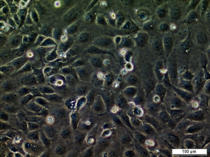
SK-MEL-3 Cell Line
SK-MEL-3 is one of a series of melanoma cell lines established from patient-derived tumour samples. This cell line is known to form tumours in immunocompromised mice.
Contributors
| Inventor | Institute |
|---|---|
| Lloyd J. Old, Germain Trempe | Memorial Sloan-Kettering Cancer Center (MSK) |
| Cat. #: | 161918 |
|---|---|
| Cancer types: | Skin cancer |
| Organism: | Human |
| Tissue: | Skin |
| Gender: | Female |
| Model: | Tumourigenic |
| Morphology: | Fibroblast |
| Growth properties: | Adherent |
| Primary citation: | Fogh et al. 1977. Journal of the National Cancer Institute. 59: 221-226. PMID: 327080. |
| Product description: | SK-MEL-3 is one of a series of melanoma cell lines established from patient-derived tumour samples. This cell line is known to form tumours in immunocompromised mice. |
|---|---|
| Gender: | Female |
| Model description: | Tumourigenic in nude mice; forms pigmented malignant melanoma |
| Products or characteristics of interest: | Karyotype: (P13) hypotetraploid to hypertetraploid with abnormalities including dicentrics, pulverizations, secondary constrictions and minutes. Isoenzymes: AK-1, 1; ES-D, 1; G6PD, B; GLO-I, 1-2; PGM1, 1-2; PGM3, 1 |
| Storage conditions: | Vapor phase of liquid nitrogen |
|---|---|
| Shipping conditions: | Dry Ice |
| Growth medium: | McCoy's 5a Medium Modified supplemented with FBS to a final concentration of 15% |
| Subculture routine: | Volumes used in this protocol are for 75 cm2 flask; proportionally reduce or increase amount of dissociation medium for culture vessels of other sizes. Remove and discard culture medium. Briefly rinse the cell layer with 0.25% (w/v) Trypsin-0.53mM EDTA solution to remove all traces of serum, which contains trypsin inhibitor. Add 2.0 to 3.0 mL of Trypsin-EDTA solution to flask and observe cells under an inverted microscope until cell layer is dispersed (usually within 5 to 15 minutes). Note: To avoid clumping do not agitate the |
| Temperature: | 37° C |
| Atmosphere: | 5% CO2 in air |
| Storage medium: | Complete growth medium supplemented with 5% (v/v) DMSO |
| Biosafety level: | 1 |
| References: |
Fogh et al. 1977. Journal of the National Cancer Institute. 59: 221-226. PMID: 327080. |
|---|
Related Tools for Cell lines
| Cat. # | Tool Name | |||||||||||||||
|---|---|---|---|---|---|---|---|---|---|---|---|---|---|---|---|---|
| 151445 | 1-7HB2 Cell Line |
Key Info
1-7HB2 Cell Line
|
View Tool | |||||||||||||
| 151503 | 1-7 CE1 Cell Line |
Key Info
1-7 CE1 Cell Line
|
View Tool | |||||||||||||
| 151579 | U-2 OS Gal4-p300 Cell Line |
Key Info
U-2 OS Gal4-p300 Cell Line
|
View Tool | |||||||||||||
| 151630 | PDVC57B Cell Line |
Key Info
PDVC57B Cell Line
|
View Tool | |||||||||||||
| 151446 | FLYRD18 Cell Line |
Key Info
FLYRD18 Cell Line
|
View Tool | |||||||||||||