Cat. #152987
HeLa mCherry-Histone H2B EGFP-Alpha Tubulin Cell Line
Cat. #: 152987
Sub-type: Continuous
Unit size: 1x10^6 cells / vial
Organism: Human
Tissue: Cervix
Model: Reporter
£575.00
This fee is applicable only for non-profit organisations. If you are a for-profit organisation or a researcher working on commercially-sponsored academic research, you will need to contact our licensing team for a commercial use license.
Contributor
Inventor: Francis Barr
Institute: University of Liverpool
Tool Details
*FOR RESEARCH USE ONLY (for other uses, please contact the licensing team)
- Name: HeLa mCherry-Histone H2B EGFP-Alpha Tubulin Cell Line
- Research fields: Cell biology;Genetics
- Tool sub type: Continuous
- Parental cell: HeLa S3
- Organism: Human
- Tissue: Cervix
- Model: Reporter
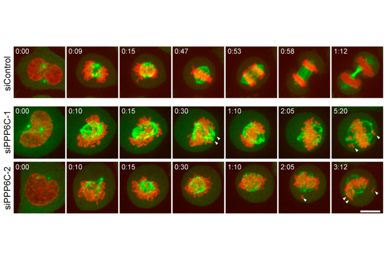
- Description: The HeLa mCherry-Histone H2B EGFP-Alpha Tubulin cell line enables live-cell imaging of chromatin (both interphase and mitotic chromosomes) and microtubule dynamics. This allows for detailed study of mitosis, spindle dynamics, cytoskeletal organisation, and cellular responses to treatments.
- Production details: The human histone H2B gene was fused to the gene encoding mCherry and Alpha Tubulin was similarly fused to EGFP. Both constructs were transfected into human HeLa cells to generate a stable line constitutively expressing H2B-mCherry and EGFP-Alpha Tubulin. The mCherry-Histone H2B fusion protein was incorporated into nucleosomes without affecting cell cycle progression.
- Recommended controls: HeLa parental line
Target Details
- Target: Histone H2B, Alpha Tubulin
Handling
- Format: Frozen
- Growth medium: DMEM, 10% FBS, 5% CO2, 37°C. Antibiotic selection for GFP and mCherry expression: 1µg/mL Puromycin, 4µg/mL Blasticidine, expression is quite stable but selecting at least every two passages is recommended.
- Unit size: 1x10^6 cells / vial
- Shipping conditions: Dry ice
Related Tools
- Related tools: HeLa EGFP-Histone H2B Cell Line
References
- Dunsch et al. 2012. J Cell Biol. 198(6):1039-1054. PMID: 22965910.
- Zeng et al. 2010. J Cell Biol. 191(7):1315-1332. PMID: 21187329.
- Bastos et al. 2010. J Cell Biol. 191(4):751-760. PMID: 21079244.


![Anti-CAR Whitlow Linker [1C3C3]](https://cancertools.org/wp-content/uploads/Figure-6-Kimble-et-al.-J-Immunother-Cancer-2025-300x322.jpg 300w, https://cancertools.org/wp-content/uploads/Figure-6-Kimble-et-al.-J-Immunother-Cancer-2025-280x300.jpg 280w, https://cancertools.org/wp-content/uploads/Figure-6-Kimble-et-al.-J-Immunother-Cancer-2025-954x1024.jpg 954w, https://cancertools.org/wp-content/uploads/Figure-6-Kimble-et-al.-J-Immunother-Cancer-2025-768x824.jpg 768w, https://cancertools.org/wp-content/uploads/Figure-6-Kimble-et-al.-J-Immunother-Cancer-2025.jpg 1193w)