Colorectal cancer (CRC), also known as bowel cancer, is the third most diagnosed cancer worldwide, with increasing incidence in younger individuals and rising mortality rates in developing countries¹. CRC arises from mutations or stable epigenetic changes in genes including adenomatous polyposis coli (APC), kirsten rat sarcoma viral proto-oncogene (KRAS), B-Raf proto-oncogene, serine/threonine kinase (BRAF) and tumour protein 53 (TP53). Only 5% of cases are linked to germline mutations, the vast majority result from somatic changes that drive tumour initiation and progression.
As research advances, effective preclinical models remain essential to understanding the molecular drivers of disease and to identify and validate new therapeutic targets.
How CancerTools.org supports CRC research and drug discovery
At CancerTools, we provide an interconnected ecosystem of CRC tools, developed by scientists, for scientists made available to the global research community to accelerate your research and preclinical drug discovery.
Our CRC portfolio can help you:
- Capture tumour heterogeneity: our portfolio includes in vitro models that mirror the complex heterogeneity of CRC, allowing you to explore tumour subtypes and variable responses with greater fidelity.
- Investigate drug responses: use our cell lines to support drug screening.
- Advance biomarker and mechanistic studies: our antibody collection provides access to highly specific, often unavailable targets, supporting target validation and biomarker discovery.
We make it easy for both academic and commercial teams to access and use these tools without unnecessary barriers.
Explore curated cell lines, antibodies and more to accelerate your colorectal cancer studies
Tumour modelling – Cell lines that capture genetic & disease diversity
Our broad collection of highly cited colorectal cancer cell lines, including patient-derived models, captures genetic and disease diversity. Offering you reliable tools for exploring tumour biology, mutation driven pathways and drug responses, supporting impactful research and drug development.

Highlights of our CRC cell line portfolio include:
- Exclusivity: 20+ unique lines, including HCT 116 derivates from Prof. Carlos Caldas and Sir Walter Bodmer’s (pictured right) pioneering models. Available only through CancerTools.org
- Genetic diversity: Lines reflecting the most frequently altered genes in CRC – KRAS, TP53, APC, BRAF, SMAD4, PIK3CA, and more, enabling studies on mutation-driven signalling and targeted therapies.
- Disease diversity: Models spanning adenocarcinomas, Dukes’ stage A-D, and metastatic origins supporting discovery and validation across heterogeneous tumours.
- Engineered variants: Explore innovative derivatives such as HCT 116 BRCA2 knockouts for DNA damage repair studies, and SW480 EGFP reporter lines for real-time monitoring of tumour cells.
Preclinical modelling
Powerful preclinical modelling with mouse models

Our curated offers powerful in vivo tools to explore CRC biology from early transformation to advanced disease and validate potential therapeutic candidates.
Highlights include:
- Cell cycle regulation models: Ideal for dissecting the roles of Fbxw7, FoxM1 & Cyclin D1 in proliferation and tumourigenesis.
- Tumour initiation & progression models: The Villin-Cre Apcfl/+ KRasG12D/+ Mouse (developed by Prof. Owen Sansom) and BRaf1 Knock-In Mouse capture early and late CRC stages, supporting research on microenvironment interactions and drug responses.
Target interrogation – Antibodies to study critical CRC pathways
Our collection of CRC antibodies provides unique access to highly specific, often unavailable targets.
Developed by leading scientists including Professors Joyce Taylor-Papadimitriou and Joy Burchel, among others, these antibodies support your research, enabling target validation, biomarker and mechanistic studies.
Highlights of our CRC antibody portfolio include:
- APC Antibodies: for probing a frequently altered tumour suppressor central to Wnt/β-catenin signalling in CRC
- MUC family antibodies: A rich panel targeting MUC1, MUC2, and MUC3, key tools for decoding mucin-related mechanisms in CRC pathogenesis
- CEACAM Antibodies: Targets including CEACAM5, CEACAM6, CEACAM8, ideal for biomarker validation and tumour profiling
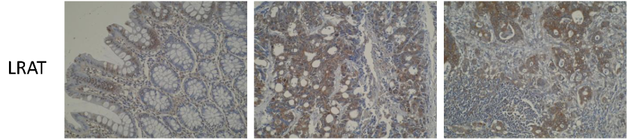
- Rare and niche targets: Featuring LRAT (Figure 12) & DRAM1 to investigate markers with emerging roles in prognosis and Cdc27 for rare, pathway-specific antibodies supporting niche areas of CRC biology
Molecular studies with vectors
The pBABE retroviral vector system continues to play a pivotal role in colorectal cancer (CRC) research thanks to its ability to support stable gene expression, sequential genetic modification, and high-titre viral delivery.

Applications in CRC:
- Modelling carcinogenesis: Recreate the stepwise genetic evolution of cancer using sequential pBABE infections³
- Oncogene characterisation: Use pBABE-puro (Figure 13) to stably overexpress candidate oncogenes in CRC cell lines, enabling long term-assessment of proliferation and invasion²
By integrating these CRC tools into your work, you are not just advancing your own research, you are contributing to a global effort to improve outcomes for people affected by colorectal cancer.
Explore the research ecosystem today and be part of the collaborative effort to transform CRC research and drug discovery.
References
- Hossain et al. 2022. Cancers (Basel). 14(7):1732. PMID: 35406504. Colorectal Cancer: A Review of Carcinogenesis, Global Epidemiology, Current Challenges, Risk Factors, Preventive and Treatment Strategies.
- Xu et al. 2014. Medicine Baltimore. 93(28):e294. PMID: 25526472. XRCC2 promotes colorectal cancer cell growth, regulates cell cycle progression, and apoptosis.
- Hahn et al. 1999. Nature. 400(6743):464-468. PMID:10440377. Creation of human tumour cells with defined genetic elements
Image credits
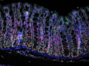
- Figure 1. Human colon. Image by Steve Bagley from the CRUK Manchester Institute and Darren Roberts from the Institute of Cancer Sciences at the University of Manchester.
- Figure 4. Bowel cancer cell. Image by Cancer Research UK London Research Institute Experimental Histopathology Unit.
- Figure 5. Migrating MDCK cells were stained for APC (green), α-tubulin (magenta), and nuclei (blue). Adapted from Boehlke et al. 2013
- Figure 6. pBABE-puro vector. Vector map created using Snapgene.
