Why the PEO series matters in ovarian cancer research
In high-grade serous ovarian cancer (HGSC), BRCA2 mutations impair DNA repair via homologous recombination (HR), creating initial sensitivity to platinum-based chemotherapy and PARP inhibitors (PARPi) (1). Resistance often emerges through secondary BRCA2 mutations that restore HR (2). This functional change is detectable via RAD51 foci formation, a widely used marker of HR competence. Studying this evolution is key to understanding and overcoming acquired drug resistance.
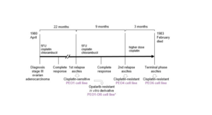
The PEO series uniquely models this trajectory. PEO1, PEO4, and PEO6 were derived from the same patient at different treatment stages. PEO1-OR, selected in vitro for olaparib resistance, extends the series by providing a model of acquired PARPi resistance, while PEO1-CDDP, an in vitro cisplatin-selected derivative of PEO1, adds a complementary platinum-resistant model. Together, these lines offer matched, mechanistically distinct models for investigating DNA repair mechanisms, drug resistance pathways, and response prediction in ovarian cancer.
Patient-matched PEO lines: modelling tumour evolution and drug resistance
| PEO1 | PEO1-OR | PEO1-CDDP | PEO4 | PEO6 | |
|---|---|---|---|---|---|
| Patient origin and treatment | From ascites of a poorly differentiated serous ovarian adenocarcinoma at first relapse, after chemotherapy (3, 4) | In vitro derivative of PEO1 selected for olaparib (PARPi) resistance (5) | In vitro derivative of PEO1 selected for cisplatin resistance (11) | From ascites of PEO1 patient, but after multiple chemotherapies (3,6) | From ascites of PEO1 patient, but at a later relapse (post-chemotherapy) (3,6) |
| BRCA2 status | Homozygous nonsense Y1655X mutation (5193C>G) causing loss of full-length BRCA2 protein (4) | Secondary mutation during selection restored the full-length HR-proficient BRCA2 protein (5) | Not reported | Secondary synonymous Y1655Y change (5193C>T) restored HR-proficient full-length BRCA2 protein (4) | Same 5193C>T silent change of PEO4, restored HR-proficient full-length BRCA2 protein (4) |
| HR capacity | Deficient with no BRCA2 protein (4) | Restored HR function and RAD51 foci–positive (5) | Not reported | Restored HR function and RAD51 foci–positive (4,5) | Expected to be RAD51 foci–positive (HR-proficient) due to its BRCA2 functionality (4) |
| Platinum sensitivity | Cisplatin-sensitive before resistance (3) | Expected to be cross-resistant to platinum due to restored BRCA2 | Cisplatin resistant (11) | Cisplatin resistant (7) | Cisplatin resistant (4) |
| PARPi sensitivity | Highly sensitive to olaparib and veliparib (8) | Selected for Olaparib resistance (5) | Resistant to AG14361 (4) and reduced sensitivity to veliparib (8) | Resistant to olaparib and veliparib (8) | |
| Tumorigenicity | Tumourigenic in immunodeficient mice (xenograft-forming) (3) | Not tested in vivo | Not tested in vivo | Tumourigenic in immunodeficient mice (xenograft-forming) (3) | Tumourigenic in immunodeficient mice (xenograft-forming) (3) |
| Model use | HRD benchmarking | PARPi resistance modelling | Platinum resistance modelling | Reverted HR-comparator | Late-stage resistance |
Abbreviations in the table: HR: homologous repair, HRD: homologous repair deficiency, PARPi: PARP inhibitor.
In addition to the core longitudinal PEO series shown above, CancerTools also distributes several related PEO-prefixed ovarian cancer cell lines (including PEO14, PEO16 and PEO23). These models share common provenance and nomenclature but do not represent sequential disease stages from a single patient. They are provided as complementary patient-derived ovarian cancer models to support a broader range of experimental applications.
References
- González-Martín A. Lancet Oncol. 2017;18(1):8–9. PMID: 27908593
- Paes Dias M, et al. Nat Rev Clin Oncol. 2021;18(12):773–791. PMID: 34285417
- Langdon et al. 1988. Cancer Research. 48(21):6166–6172. PMID: 3167863
- Sakai et al. 2009. Cancer Research. 69(16):6381–6386. PMID: 19654294
- Biegala et al. 2023. Cells. 12(7), 1038. PMID: 37048111
- Greenwood et al. Clin Cancer Res. 2019. 25(8):2471-2482. PMID: 30651275
- Zhihong et al. 2016. Cancer Lett. 19;373(1):36–44. PMID: 26801746
- Stukova et al. 2015. J Inorg Biochem. 149:45-8. PMID: 26021697
- Ng CKY et al. 2012. J. Pathol. 226(5):703-12. PMID: 22183581.
- Meijer TG et al. 2024. Cancers (Basel). 16(4):741. PMID: 38398132.
- Macleod et al. 2005. Cancer Res. 65(15):6789-800. PMID: 16061661
