C10 Colorectal Cell Line
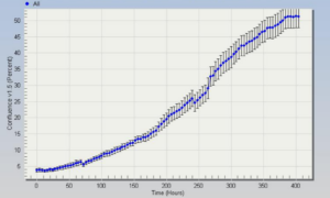
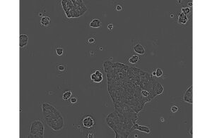
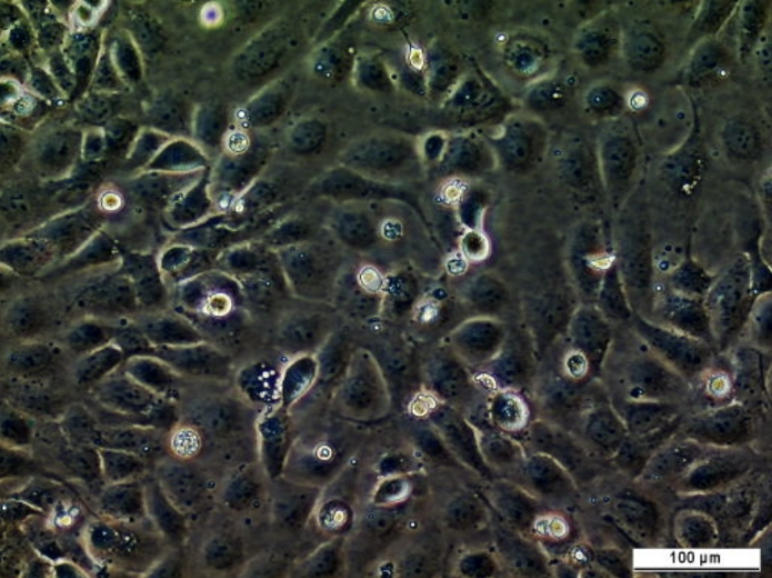
This C10 colorectal cell line was established from surgically resected colorectal adenocarcinomas. Research Area, Cancer, Drug Discovery & Development.
Images
View Gallery

This C10 colorectal cell line was established from surgically resected colorectal adenocarcinomas. Research Area, Cancer, Drug Discovery & Development.

Please note we may take up to three days to respond to your enquiry.
CancerTools.org uses the contact information provided to respond to you about our research tools and service. For more information please review our privacy policy.