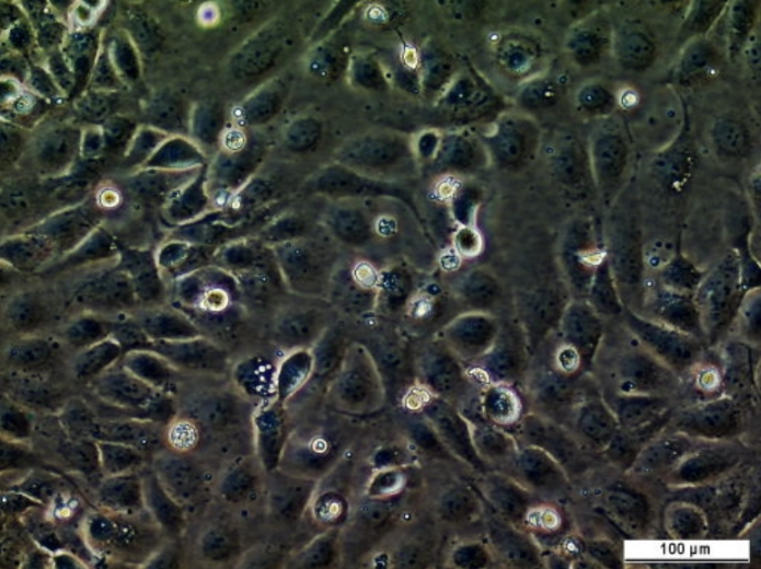
#161916

SK-MEL-24 Cell Line

Cat. #161916

SK-MEL-24 Cell Line

Cat. #: 161916

Availability: 8-10 weeks

Organism: Human

Tissue: Skin

Model: Tumourigenic

£575.00

This fee is applicable only for non-profit organisations. If you are a for-profit organisation or a researcher working on commercially-sponsored academic research, you will need to contact our licensing team for a commercial use license.

Contributor

Inventor: Lloyd J. Old, Toshitada Takahashi

Institute: Memorial Sloan-Kettering Cancer Center (MSK)

Primary Citation: Carey et al. 1976. Proceedings of the National Academy of Sciences. 73: 3278-3282. PMID: 1067619.

Tool Details
Target Details
Applications
Handling
References

Tool Details

*FOR RESEARCH USE ONLY (for other uses, please contact the licensing team)

  • Name: SK-MEL-24 Cell Line
  • Cancer: Skin cancer
  • Cancers detailed: Melanoma
  • Organism: Human
  • Gender: Male
  • Tissue: Skin
  • Donor: This cell line was established from a metastatic site (lymph node) in a 67-year-old Caucasian male with malignant melanoma.
  • Morphology: Stellate
  • Growth properties: Adherent
  • Model: Tumourigenic
  • Model description: Tumourigenic; forms malignant melanoma
  • Products or characteristics of interest: Isoenzymes: AK-1, 1; ES-D, 1; G6PD, B; GLO-I, 1-2; Me-2, 2; PGM1, 1; PGM3, 2
  • Description: K-MEL-24 is one of a series of melanoma cell lines established from patient-derived tumour samples. This cell line expresses wildtype B-Raf and wildtype N-Ras.
  • Application: 3D cell culture
  • Biosafety level: 1

Target Details

  • Target: Antigen expression: Blood Type O; Rh+; HLA A1, A2, B12, B14, Cw5

Applications

  • Application: 3D cell culture

Handling

  • Growth medium: EMEM supplemented with FBS to a final concentration of 15%
  • Temperature: 37° C
  • Atmosphere: 5% CO2 in air
  • Shipping conditions: Dry Ice
  • Storage medium: Complete growth medium supplemented with 5% (v/v) DMSO
  • Storage conditions: Vapor phase of liquid nitrogen
  • Subculture routine: Remove and discard culture medium. Briefly rinse the cell layer with 0.25% (w/v) Trypsin- 0.53 mM EDTA solution to remove all traces of serum that contains trypsin inhibitor. Add 2.0 to 3.0 mL of Trypsin-EDTA solution to flask and observe cells under an inverted microscope until cell layer is dispersed (usually within 5 to 15 minutes). Note: To avoid clumping do not agitate the cells by hitting or shaking
  • Str profiling: Amelogenin: X,YCSF1PO: 11D13S317: 12,13D16S539: 9,12D5S818: 9,13D7S820: 9,12TH01: 6,9TPOX: 11vWA: 15D3S1358: 15D21S11: 29,31.2D18S51: 16Penta_E: 11,12Penta_D: 9D8S1179: 14,15FGA: 22,23D19S433: 13,14D2S1338: 23

References

  • Xing et al. 2012. Oncogene. 31(4):446-457. PMID: 21725359.

Tool enquiry

Please ensure you use your organisation email address rather than personal where possible, as this helps us locate your organisation in our system faster.

Please note we may take up to three days to respond to your enquiry.

CancerTools.org uses the contact information provided to respond to you about our research tools and service. For more information please review our privacy policy.