Explore a portfolio of research models, including cell lines and antibodies, developed by ovarian cancer researchers worldwide and shared to accelerate discovery. Our tools are available to academic researchers via our website and to commercial partners via our dedicated licensing support. Our ovarian cancer research tools at a glance:
- 80+ ovarian cancer cell lines
- 50+ ovarian cancer antibodies
The challenge in ovarian cancer research
Ovarian cancer is one of the most prevalent gynaecological malignancies worldwide, accounting for 3.7% of all cancer cases and 4.7% of cancer deaths in 2020. 1, 2 .
Its most common variant, epithelial ovarian cancer (EOC), includes five genetically distinct subtypes with unique therapeutic responses:
- High-Grade Serous Carcinoma (HGSC) [~75% of cases]
- Endometrioid carcinoma
- Clear cell carcinoma
- Low-grade serous carcinoma
- Mucinous carcinoma
This complexity means standard models are often insufficient. To meet this need, CancerTools.org provides a comprehensive set of ovarian cancer research models for disease modelling and preclinical drug discovery that capture diversity, resistance, and clinical context.
Our perspective: visual insights into drug resistance
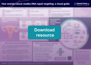
At CancerTools.org, we not only provide access to ovarian cancer models, we also share insights that advance understanding of complex challenges such as drug resistance.
Our infographic, “How ovarian cancer evades DNA repair targeting”, offers a visual overview of the mechanisms that shape therapy response and resistance.
Get the complete infographic to explore how DNA repair pathways influence ovarian cancer research and discover the models that make this possible.
Our ovarian cancer research portfolio
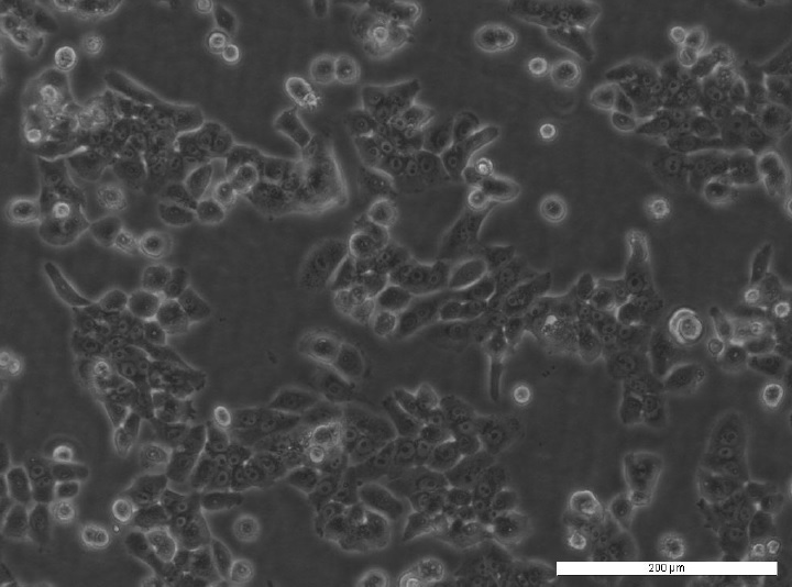
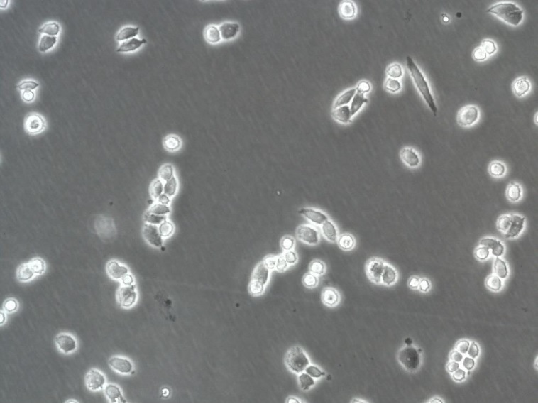
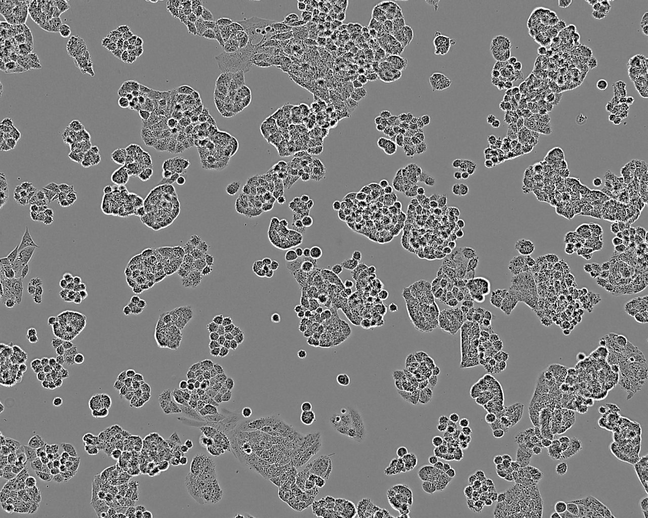
Ovarian cancer cell lines for diverse applications
Ovarian cancer’s complexity, from subtype heterogeneity to drug resistance, demands a layered approach. Our ovarian cancer cell line portfolio helps researchers to recreate this spectrum in the lab for:
- Mechanistic studies
- Drug discovery
- Resistance profiling
- Tumour microenvironment research
Models for studying drug resistance in ovarian cancer
Drug resistance is one of the greatest challenges in ovarian cancer treatment. Our portfolio offers human cell lines that enable researchers to track this process over time, from patient-derived longitudinal systems to engineered resistant derivatives and clinically relevant panels.
PEO Series: longitudinal resistance model
The PEO series comprises matched HGSC models from the same patient, including BRCA2-mutant, therapy-sensitive PEO1 and platinum-resistant derivatives such as PEO4 and PEO6.
These lines uniquely map the evolution of DNA repair defects and therapy response and are also used to study hormone receptor biology, such as oestrogen response.
A2780-derived resistance models
Derivatives of the A2780, parental line provide reproducible systems for studying resistance across key drug classes:
- Cisplatin: A2780cis and PEO1-CDDP — modelling platinum resistance, the most common clinical hurdle in ovarian cancer.
- Adriamycin: A2780ADR — complementary model for anthracycline resistance.
- Paclitaxel: A2780 PTX isogenic series (PTX4–128) — stepwise taxane resistance with graded collateral cisplatin sensitivity, distinct from platinum and anthracycline lines, enabling comparative studies.
Platinum-exposed HGSC resistance models

This platinum-resistant HGSC panel was developed from well-established human parental lines, including OVCAR4, Ovsaho, and Cov318, to enable comparative studies of resistance evolution following cisplatin or carboplatin exposure.
Rather than representing a single resistant state, the series captures biological and phenotypic heterogeneity associated with relapse, with models displaying distinct growth and invasive behaviours in vivo, from aggressive ascites formation to slower metastatic spread. These features are consistent with transcriptional reprogramming associated with platinum-resistant ovarian cancer, including extracellular matrix–associated pathways (Hoare et al., 2022).
Individual models can be used independently, while their value is maximised when studied together as a coordinated system to distinguish shared adaptive programmes from context-specific resistance mechanisms. The panel complements other ovarian cancer resistance models, such as A2780 derivatives, by supporting questions focused on relapse biology, comparative resistance trajectories, and tumour behaviour beyond drug sensitivity alone.
A2780: a foundational ovarian cancer line
One of the most widely used ovarian cancer cell lines, A2780, is a cornerstone for toxicity testing and cancer genetics research.
It can induce tumours in immunocompromised mice, making it a reliable in vivo model as well as a critical reference for its many resistant derivatives.
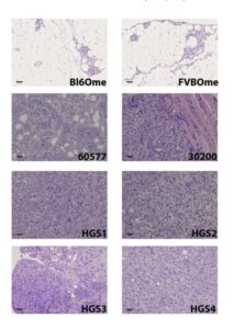
Murine HGSC models to investigate tumour–immune interactions
Developed in immunocompetent mouse models HGS 1-4 recreate many hallmarks of human high-grade serous ovarian cancer, from genetic drivers to metastatic spread.
Used in vivo, they provide a rare opportunity to investigate tumour–immune interactions and therapy response in a biologically relevant setting.
OCM series: patient tumour fractions
The OCM series represents highly purified HGSOC tumour fractions with strong proliferative potential, analysed at early passage to minimise culture drift.
These models preserve the molecular features of the original tumours, are clinically annotated, and are included in public datasets such as multi-omics and single-cell karyotyping.
OCM cultures enable reliable drug-sensitivity studies of tumour cells without extensive passaging or genetic drift and can also generate PDX models for in vivo evaluation of new therapies.
Patient-derived ovarian cancer lines
Capturing the complexity of the tumour microenvironment is essential for preclinical models of ovarian cancer. Our patient-derived collections reflect this diversity, spanning different tumour sites, histotypes, and treatment stages while retaining the molecular characteristics of the original disease.
- CCSP series (6 lines): tumourigenic cell lines derived from malignant ascites in patients with clear cell adenocarcinoma, providing a rare and aggressive OEC subtype for modelling
- OV cell lines (14 lines): ascites-derived models representing primarily high-grade serous carcinoma
- TOV cell lines (18 lines): derived from ovary, endometrium, and fallopian tube tumours, capturing the spectrum of EOC histotypes
Antibodies for ovarian cancer research
Our portfolio includes widely used antibodies against key proteins in ovarian cancer pathways, supporting studies from DNA repair to hormone signalling and tumour antigen biology.
- DNA repair proteins, such as PALB2, a central scaffolding factor in homologous recombination repair with BRCA1/2.
- Hormone receptors, such as oestrogen receptor β, relevant for hormone-dependent ovarian cancer biology.
- Tumour-associated antigens, such as MUC1, frequently overexpressed and abnormally glycosylated in late-stage epithelial ovarian cancer.
