#153570

MET4 SCC Cell Line

Cat. #153570

MET4 SCC Cell Line

Cat. #: 153570

Organism: Human

Tissue: Skin

Model: Tumour line

This fee is applicable only for non-profit organisations. If you are a for-profit organisation or a researcher working on commercially-sponsored academic research, you will need to contact our licensing team for a commercial use license.

Contributor

Inventor: Irene Leigh

Institute: Queen Mary University of London

Tool Details
Target Details
Handling
Related Tools
References

Tool Details

*FOR RESEARCH USE ONLY (for other uses, please contact the licensing team)

  • Name: MET4 SCC Cell Line
  • Alternate name: SCC, Keratinocyte
  • Cancer type filter: Skin cancer
  • Organism: Human
  • Tissue: Skin
  • Donor: Single immunosuppressed patient, from a primary invasive SCC on the back of the hand that subsequently recurred locally twice
  • Model: Tumour line
  • Description: 
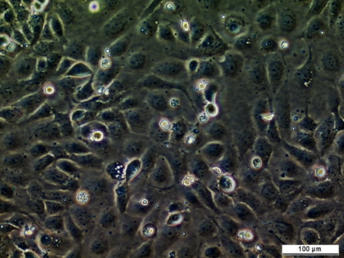
Keratinocyte cell line, representing the ultimate metastatic stage in a SCC cancerous transformation. Background and Research Application MET4 is derived from a from metastatic SCC within left axillary lymph nodes that were biopsied following the third excision. The cell lines originated as a primary tumour on the left hand. Histology of the original tumour showed an acantholytic squamous cell carcinoma. MET4 has been used to understand aggressive metastatic SCCs and is an important cell line for this specific stage in SCC cancer progression. MET4 is the fourth cell line in a unique series of epidermal cell lines representing different stages of malignant transformation. These cell lines were derived from a single, adult, immunosuppressed individual. Four keratinocyte lines (PM1-4) established from forehead skin are here compared with 4 squamous cell carcinoma (SCC) lines MET2 derived respectively from a primary cutaneous tumour, two local recurrences and a distant metastasis of invasive SCC. Despite altered growth properties, the PM lines retained many features of normal keratinocytes including keratin phenotype, differentiation capacity and non-tumorigenicity in athymic mice. In contrast, from early passage, the MET lines displayed markedly reduced growth requirements, abnormal differentiation, aberrant K18 expression and tumorigenicity in athymic mice. The abnormal keratin profile of individual MET lines closely reflected the keratin phenotype of the tumour of origin. Although unusual HPV types were identified in the original tissue, there was no evidence of persistent virus within any cell line and it appears that HPV is not critical for maintenance of the immortal phenotype. The PM lines were distinctly different from invasive SCC lines and are likely to be useful in studies focusing on mutations that are important in early neoplastic progression. The SCC series represent primary, recurrent and metastatic carcinoma. Availability of such a series from the same individual will facilitate genetic analysis of the malignant process.
  • Production details: MET4 was derived from a metastatic SCC within the left auxillary lymph nodes that were biopsied
  • Biosafety level: 1
  • Additional notes: Difference Between Clones All derived from the same individual, but from varying stages in progression of an SCC: invasive, recurrent and, subsequently metastatic. Keratinocyte lines MET1, MET2, MET3 and MET4 were derived, respectively, from an SCC excised from the back of the left hand (MET1), from 2 recurrent SCC arising at the same site in rapid succession (MET2 and 3)and from metastatic SCC within left axillary lymph nodes biopsied following the third excision (MET4).

Target Details

  • Target: Squamous Cell Carcinoma line

Handling

  • Format: Frozen
  • Growth medium: Keratinocyte cell culture media supplemented with 10ng/ml EGF (Long term growth), DMEM/5% serum (Banking purposes)
  • Shipping conditions: Dry ice
  • Storage conditions: Liquid Nitrogen

Related Tools

  • Related tools: MET1 SCC Cell Line ; MET2 SCC Cell Line

References

  • Inman et al. 2018. Nat Commun. 9(1):3667. PMID: 30202019.
  • Abikhair Burgo et al. 2018. JCI Insight. 3(17):. PMID: 30185657.
  • Mekhdjian et al. 2017. Mol Biol Cell. 28(11):1467-1488. PMID: 28381423.
  • Proby et al. 2000. Exp Dermatol. 9(2):104-17. PMID: 10772384.

Tool enquiry

Please ensure you use your organisation email address rather than personal where possible, as this helps us locate your organisation in our system faster.

Please note we may take up to three days to respond to your enquiry.

CancerTools.org uses the contact information provided to respond to you about our research tools and service. For more information please review our privacy policy.