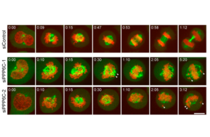
HeLa mCherry-Histone H2B EGFP-Alpha Tubulin Cell Line
The human histone H2B gene was fused to the gene encoding mCherry and Alpha Tubulin was similarly fused to EGFP.
Images
View Gallery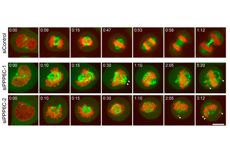
The human histone H2B gene was fused to the gene encoding mCherry and Alpha Tubulin was similarly fused to EGFP.

Please note we may take up to three days to respond to your enquiry.
CancerTools.org uses the contact information provided to respond to you about our research tools and service. For more information please review our privacy policy.