Home » Resources » Tumour Immunology Antibodies
Tumour immunology studies the complex interactions between cancer cells and the immune system.
Research in this field aims to uncover the mechanisms by which the immune system inhibits cancer growth via anti-tumour responses and promotes tumour progression through immunosuppression.
Research in tumour immunology also aims to identify new ways of harnessing the power of the immune system to tackle cancer, including developing new immunotherapy treatments, such as immune checkpoint and adoptive T cell transfer therapies.
CancerTools.org has established the first-of-its-kind non-profit antibody collection where 2000+ antibodies developed by scientists globally have been curated in a single online collection for easy access. The antibodies and other research tools below are a selection of the research tools available in key research areas in tumour immunology including:
Immunophenotyping
Analysing immune cells has become a key part of any research in tumour immunology. The CancerTools.org collection includes:
- antibodies for studying innate immunity including those directed at macrophages and dendritic cells;
- For research in adaptive immunity, we have antibodies for T cells, T cell
receptors, and markers of different subsets of T cells like CD8 (cytotoxic) and CD4 (helper T cells); - antibodies for proteins involved in antigen presentation, e.g., MHC/HLA; as well as
- an extensive range of CD markers.
Image: Fluorescent staining of dendritic cells
Immune checkpoints
Harnessing immune checkpoint inhibitors as immunotherapeutic agents represented a breakthrough in cancer immunotherapy. Immune checkpoints are regulators of immune activation that maintain self-tolerance by controlling the duration and extent of the immune response.
Cancer cells exploit these regulators by upregulating immune checkpoint proteins like PD-L1 and CTLA4 which negatively regulate the immune response, thus allowing tumour cells to evade the immune system.
The immunology collection features:
- antibodies recognising key co-inhibitory molecules in immune checkpoint control, such as PD-1, PDL-1, and CTLA-4; as well as
- co-stimulatory molecules, like CD40 and CD70.
Image: Immune checkpoint interactions between a T cell and
a cancer cell. Created with BioRender.com
Tumour microenvironment
The tumour microenvironment (TME) is comprised of blood vessels, immune cells, stromal cells, signalling molecules and extracellular matrix surrounding the tumour. Its composition varies between tumour types, and it plays an active role in cancer progression. Better understanding of the TME regulation by the immune system is key to harness anti-tumour immune responses.
The CancerTools.org antibodies can be used to study key processes occurring in the TME, such as: vascularisation, hypoxia, extracellular matrix remodelling, and inflammation. Targets included in the collection are:
- chemokines;
- HIF1α;
- VEGF; and
- matrix metalloproteinases (MMPs).
The CancerTools.org collection also has a vector that can be used to identify cells in the metastatic niche and cell lines that can be used as models of the ovarian TME.
Image:Tumour microenvironment, including cancer cells, immune cells, fibroblasts, inflammatory mediators, and vascular components. Created with BioRender.com
Inflammation
Inflammation is the biological response to stimuli by invading pathogens or endogenous signals, e.g., damaged cells, that results in tissue repair or pathology. It is considered the driving factor in many diseases, including atherosclerosis, autoimmunity, infections, and cancer.
The collection offers antibodies:
- against biomarkers of inflammation such as c-reactive protein, fibrinogen-like protein 1 and myeloperoxidase;
- for key adhesion molecules and chemokines, enabling the evaluation of leukocyte infiltration and activation; as well as
- for immune cells mediating inflammation such as neutrophils, macrophages, mast cells and dendritic cells.
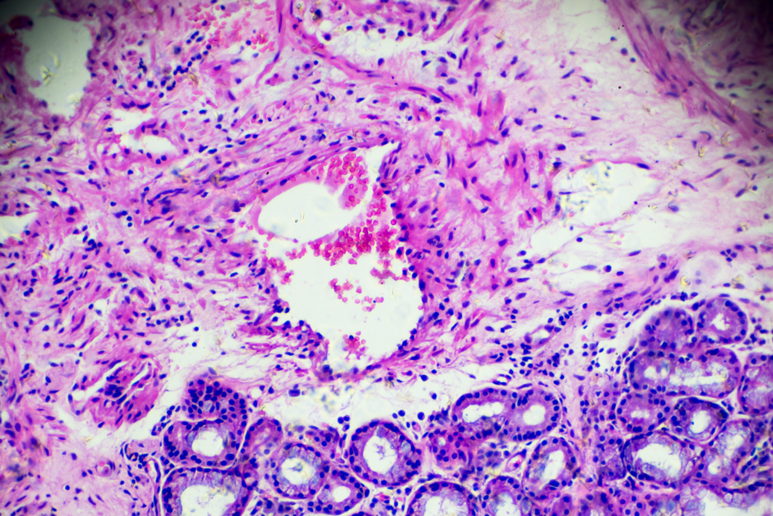
Image:Inflammatory cells in histopathological sample.
Tumour-intrinsic signalling pathways
Tumour-intrinsic signalling pathways are considered oncogenic and play a key role in regulating the immunosuppressive tumour microenvironment and tumour immune escape.
These signalling pathways include STAT3, β-Catenin, PI3K/PTEN/AKT/mTOR, p53, NF-κB, RAS/RAF/MAPK, and KRAS/MYC. They can drive effective immunocyte exclusion and disfunction, as well as recruitment and differentiation of immunosuppressive cells in various tumour types.
The antibody collection targets a wide range of molecules involved in these signalling pathways, including AKT1, RAF, MYC and p53.
Image: Fluorescent staining of mammary tumour in MDA-MB-231-cell line derived orthotopic model. Green staining is for Omomyc, red for Ki67, and blue for DAPI. Photo credit: Laura Soucek.
Epigenetics
Epigenetic mechanisms, including DNA methylation, histone post-translational modifications, and chromatin structure regulation, are often used by tumours to escape immune restriction.
Epigenetic targets of the antibody collection include:
- DNA methylation, such as 5-hydroxymethylcytosine and
anti-N6-methyladenosine; as well as - ubiquitination and other post-translational modifications, such as O-GlcNAcylation.
Image: Histone post-translational modifications. Created with BioRender.com
Read our latest on tumour immunology


