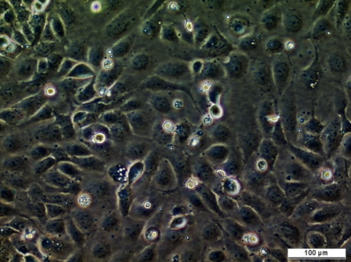
Cat. #161924
Calu-6 Cell Line
Cat. #: 161924
Availability: 8-10 weeks
Organism: Human
Tissue: Unknown
Model: Tumourigenic
£575.00
This fee is applicable only for non-profit organisations. If you are a for-profit organisation or a researcher working on commercially-sponsored academic research, you will need to contact our licensing team for a commercial use license.
Contributor
Inventor: Jorgen Fogh
Institute: Memorial Sloan-Kettering Cancer Center (MSK)
Primary Citation: Wright et al. 1981. Journal of the National Cancer Institute. 66:239-247. PMID: 6936474.
Tool Details
*FOR RESEARCH USE ONLY (for other uses, please contact the licensing team)
- Name: Calu-6 Cell Line
- Cancer type: Lung cancer
- Organism: Human
- Gender: Female
- Tissue: Unknown
- Donor: This cell line was established in 1971 from lung metastasis in lung adenocarcinoma
- Morphology: Epithelial
- Growth properties: Adherent
- Model: Tumourigenic
- Model description: Tumourigenic in nude mice; forms poorly differentiated carcinoma
- Products or characteristics of interest: Karyotype: The stemline chromosome number is hypotriploid and the 2S component occurred at 5.8%. Modal chromosome number is 59. Fourteen marker chromosomes (constitutive) were common to most S metaphases. No Y chromosome was detected in the QM stained preparation. Isoenzymes: AK-1, 1; ES-D, 1; G6PD, B; GLO-I, 2; Me-2, 1; PGM1, 2; PGM3, 1
- Description: This cell line was established in 1971 from lung metastasis in lung adenocarcinoma. CaLu-6 is a human lung adenocarcinoma cell line.
- Application: 3D cell culture; Cancer research
- Biosafety level: 1
Applications
- Application: 3D cell culture; Cancer research
Handling
- Growth medium: EMEM supplemented with FBS to a final concentration of 10%
- Temperature: 37° C
- Atmosphere: 5% CO2 in air
- Shipping conditions: Dry Ice
- Storage medium: Complete growth medium supplemented with 5% (v/v) DMSO
- Storage conditions: Vapor phase of liquid nitrogen
- Str profiling: Amelogenin: XCSF1PO: 12D13S317: 11D16S539: 13D5S818: 11D7S820: 10TH01: 9TPOX: 8vWA: 17D3S1358: 16D21S11: 31D18S51: 12,16Penta_E: 5,14Penta_D: 13D8S1179: 10,14FGA: 22D19S433: 13D2S1338: 17,26
References
- Wright et al. 1981. Journal of the National Cancer Institute. 66:239-247. PMID: 6936474.
![Anti-CAR Whitlow Linker [1C3C3]](https://cancertools.org/wp-content/uploads/Figure-6-Kimble-et-al.-J-Immunother-Cancer-2025-300x322.jpg 300w, https://cancertools.org/wp-content/uploads/Figure-6-Kimble-et-al.-J-Immunother-Cancer-2025-280x300.jpg 280w, https://cancertools.org/wp-content/uploads/Figure-6-Kimble-et-al.-J-Immunother-Cancer-2025-954x1024.jpg 954w, https://cancertools.org/wp-content/uploads/Figure-6-Kimble-et-al.-J-Immunother-Cancer-2025-768x824.jpg 768w, https://cancertools.org/wp-content/uploads/Figure-6-Kimble-et-al.-J-Immunother-Cancer-2025.jpg 1193w)

![Anti-CAR Whitlow Linker [1B4A1]](https://cancertools.org/wp-content/uploads/Figure-5-Kimble-et-al.-J-Immunother-Cancer-2025-300x396.jpg 300w, https://cancertools.org/wp-content/uploads/Figure-5-Kimble-et-al.-J-Immunother-Cancer-2025-227x300.jpg 227w, https://cancertools.org/wp-content/uploads/Figure-5-Kimble-et-al.-J-Immunother-Cancer-2025-776x1024.jpg 776w, https://cancertools.org/wp-content/uploads/Figure-5-Kimble-et-al.-J-Immunother-Cancer-2025-768x1013.jpg 768w, https://cancertools.org/wp-content/uploads/Figure-5-Kimble-et-al.-J-Immunother-Cancer-2025.jpg 970w)


