Cat. #152934
Anti-deltaEGFR [DH8.3] mAb
Cat. #: 152934
Sub-type: Primary antibody
Unit size: 100 ug
Availability: 3-4 weeks
Target: Epidermal growth factor receptor, truncated EGF receptor (RGFR typeIII/EGFRvIII/deltaEGFR). Recognises an EGFR with truncated extracellular domain, present on human tumours.
Class: Monoclonal
Application: ELISA ; FACS ; IHC ; IP ; WB
Reactivity: Human
Host: Mouse
£300.00
This fee is applicable only for non-profit organisations. If you are a for-profit organisation or a researcher working on commercially-sponsored academic research, you will need to contact our licensing team for a commercial use license.
Contributor
Inventor: Bill Gullick
Institute: Cancer Research UK, London Research Institute: Lincoln's Inn Fields
Primary Citation: Hills et al. 1995. Int J Cancer. 63(4):537-43. PMID: 7591264.
Tool Details
*FOR RESEARCH USE ONLY (for other uses, please contact the licensing team)
- Name: Anti-deltaEGFR [DH8.3] mAb
- Alternate name: EGFRvIII, Avian erythroblastic leukemia viral (v erb b) oncogene homolog, Cell growth inhibiting protein 4, Cell proliferation inducing protein 61, EGF R, EGFR, Epidermal growth factor receptor (avian erythroblastic leukemia viral (v erb b) oncogene homolog), Epidermal growth factor receptor (erythroblastic leukemia viral (v erb b) oncogene homolog avian), Epidermal growth factor receptor, erb-b2 receptor tyrosine kinase 1, ERBB, ERBB1, Errp, HER1, mENA, NISBD2, Oncogen ERBB, PIG61, Proto-onc...
- Cancer: Neurological cancer
- Cancers detailed: Brain;Glioblastoma;Glioblastoma Multiforme
- Research fields: Cancer;Cell signaling and signal transduction
- Clone: DH8.3
- Tool sub type: Primary antibody
- Class: Monoclonal
- Conjugation: Unconjugated
- Cell signalling pathway: EGFR
- Reactivity: Human
- Host: Mouse
- Application: ELISA ; FACS ; IHC ; IP ; WB
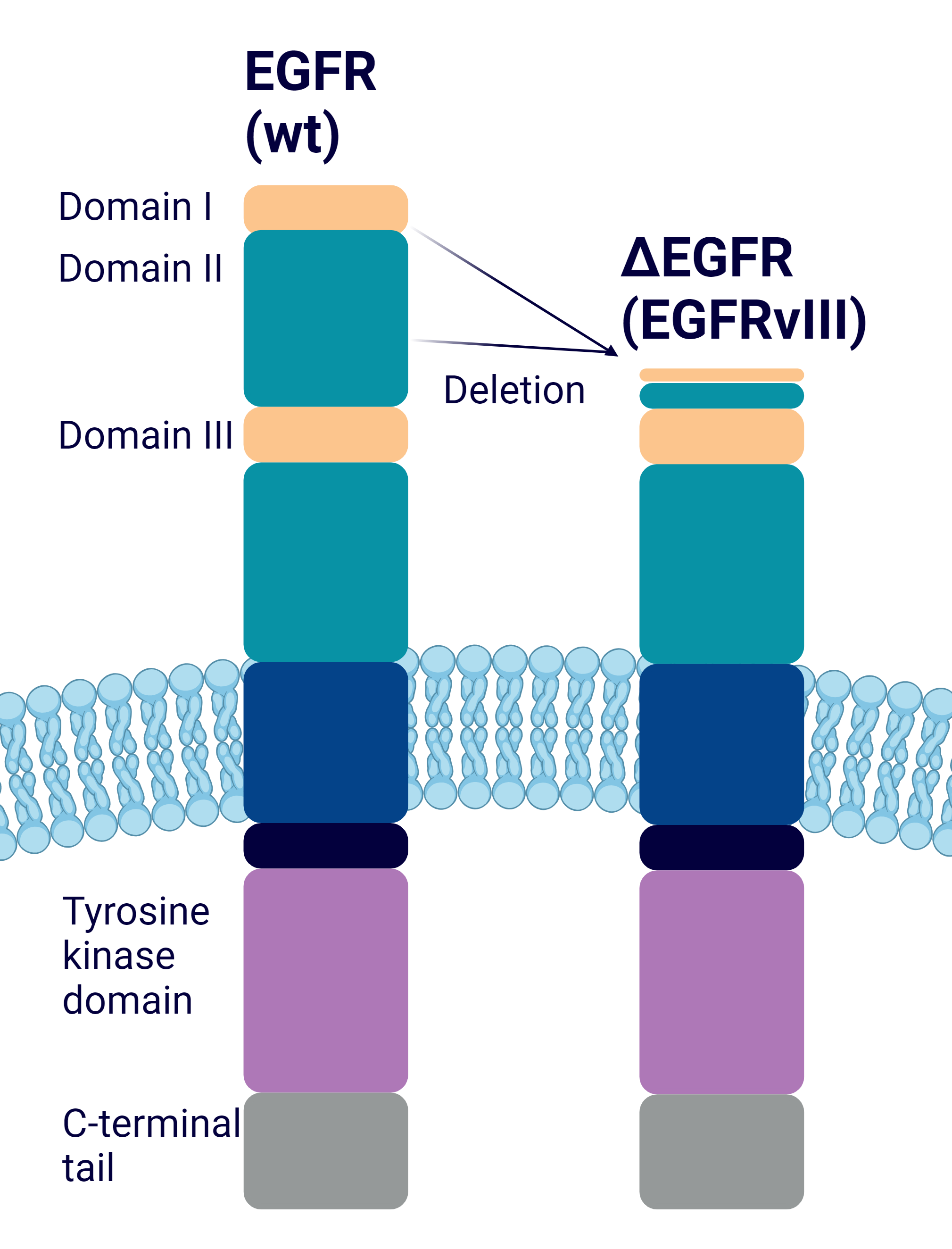
- Description: Monoclonal antibody directed against ΔEGFR, a mutated form of EGFR, typically found in brain tumours, also known as EGFRvIII. This antibody has use within radiolabelled tumour imaging and potentially therapeutic targeting against ΔEGFR tumours. Background and Research Applications: Epidermal growth factor (EGFR) has attracted considerable attention as a target for cancer therapy. Wild-type EGFR is amplified in a number of cancers, and several mutant forms of the EGFR coding gene have been found. ΔEGFR is the most common mutation, with a deletion of exons 2-7 in the external domain of wt-EGFR, generating a truncated form of EGFR. Anti-ΔEGFR [DH8.3] is raised to a synthetic peptide that recognises the junctional region of the ΔEGFR receptor. Points of Interest Anti-ΔEGFR [DH8.3] does not recognise the natural, wild-type form of EGFR receptor, so therefore does not cross react with the full-length receptor. It only binds cells expressing the mutant receptor. This antibody can be used in tumour imaging, with radiolabelled antibody DH8.3. This antibody has successfully targeted tumours expressing DH8.3 antigen in nude mice. This antibody recognises ΔEGFR in both denatured and native states.
- Immunogen: Synthetic peptide corresponding to the junctional region of the truncated receptor LEEKKGNYVVTDHC,conjugated to keyhole limpet haemcyanin.
- Immunogen uniprot id: P00533
- Isotype: IgG1 kappa
- Myeloma used: NS0
Target Details
- Target: Epidermal growth factor receptor, truncated EGF receptor (RGFR typeIII/EGFRvIII/deltaEGFR). Recognises an EGFR with truncated extracellular domain, present on human tumours.
- Target background: ΔEGFR is the most common alteration observed in glioblastomas. It has a deletion of exons 2 to 7 in the external domain of wt-EGFR, generating a truncated form of EGFR. This truncated EGFRvIII is constitutively active. In this genetic rearrangement where exon 1 is spliced to exon 8, resulting in the loss of 801 bp from the mature mRNA. This corresponds to a deletion of 267 amino acids in the receptor's extracellular domain. The structure of the receptor is then unable to bind ligand, yet is constitutively active, enhancing tumorigenesis due to impaired internalisation and degradation.
Applications
- Application: ELISA ; FACS ; IHC ; IP ; WB
Handling
- Format: Liquid
- Concentration: 1 mg/ml
- Unit size: 100 ug
- Storage buffer: PBS with 0.02% azide
- Storage conditions: Store at -20° C frozen. Avoid repeated freeze / thaw cycles
- Shipping conditions: Dry ice
References
- Genßler et al. 2016. Oncoimmunology. 5(4):e1119354. PMID: 27141401
- Feng et al. 2014. J Clin Invest. 124(9):3741-56. PMID: 25061874
- Feng et al. 2012. Proc Natl Acad Sci U S A. 109(8):3018-23. PMID: 22323579
- Lammering et al. 2004. Clin Cancer Res. 10(19):6732-43. PMID: 15475464
- Nishikawa et al. 2004. Brain Tumor Pathol. 21(2):53-6. PMID: 15700833
- Jungbluth et al. 2003. Proc Natl Acad Sci U S A. 100(2):639-44. PMID: 12515857
- Johns et al. 2002. Int J Cancer. 98(3):398-408. PMID: 11920591
- Hills et al. 1995. Int J Cancer. 63(4):537-43. PMID: 7591264


