HCI-012 breast cancer PDX
HCI-012 PDX: Human breast cancer-derived xenograft retains high fidelity to original tumour. Valuable resources for drug discovery and precision oncology.
Contributors
| Inventor | Institute |
|---|---|
| Alana L Welm, Yi-Chun Lin, Yoko Sakata DeRose | The University of Utah Research Foundation |
| Cat. #: | 162080 |
|---|---|
| Cancer types: | Breast cancer |
| Cancer Subtype: | Infiltrating Ductal Carcinoma |
| Gender: | Female |
| Cancer Stage or Grade: | Stage IV |
| Biopsy Site: | Pleural Effusion Fluid |
| Patient ethnicity: | Caucasian |
| Treatment History: | Pretreated: Patient had undergone radiation therapy to neck and lung (2009) and had systemic treatment of nab-paclitaxel; 5-fluorouracil; cyclophosphamide; aldesleukin/proleukin; interleukin 2; epirubicin, leucovarin, methotrexate; letrozole (2006 – 2009); doxorubicin (2009); capecitabine (2009), trastuzumab, vinerolbine (2009), capecitibine and lapatinab (2009) prior to sample collection |
| Primary citation: | Guillen, et al. 2022. Nature Cancer. Feb; 3(2):232-250. PMID: 35221336 |
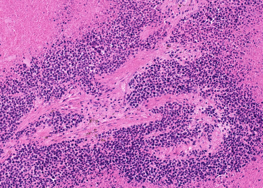
| Product description: | Human breast cancer patient-derived xenograft (PDX) model that retains high fidelity to original tumour, including spatial structure, intratumour heterogeneity, genomic features, tumour growth rate, metastatic patterns, and drug responses. These highly translatable PDX models can be used to more accurately assess therapeutic efficacy and predict patient responses in preclinical drug validation studies than traditional models. This panel of PDX models includes some of the deadliest forms of breast cancer such as drug-resistant, metastatic tumours, and endocrine-resistant estrogen receptor-positive (ER+) and human epidermal growth factor receptor positive (HER2+) tumours. Sample collected in 2009 from pleural effusion of Caucasian female, age 51 at time of collection with a primary diagnosis of IDC; 2000; recurrences in 2007 and 2008. Patient was a smoker for 14 years, and experienced clinical metastasis to the lymph node and pericardium. Patient had undergone radiation therapy to neck and lung (2009) and had systemic treatment of nab-paclitaxel; 5-fluorouracil; cyclophosphamide; aldesleukin/proleukin; interleukin 2; epirubicin, leucovarin, methotrexate; letrozole (2006 – 2009); doxorubicin (2009); capecitabine (2009), trastuzumab, vinerolbine (2009), capecitibine and lapatinab (2009) prior to sample collection. Patient characteristics were as follows – ER status: negative, PR status: negative, HER2 status: primary tumour was positive and amplified; PE not tested. PDX characteristics were as follows – ER status: negative, PR status: negative, HER2 status: positive, mixed. PDX information: PAM50 subtype is luminal B/HER2-enriched, PTEN negative by IHC, and shows metastasis to lung, thyumus, ovary, kidney, liver and lymph node. |
|---|
| Initial handling information: | Implant into the cleared inguinal mammary fat pad of female Immune-compromised mice (NOD.SCID) and NGS. Time to grow to 2cm: 4 months (both) |
|---|---|
| Additional notes: | Additional Information on PDX establishment: https://www.nature.com/articles/s43018-022-00337-6/figures/9 |
| Mice Passaged: | Yes |
|---|---|
| Engraftment Site: | Cleared mammary fat pad |
| Sample Type: | Suspension in Matrigel |
| Host Strain: | Immunocompromised mice NOD scid gamma (NSG) Jackson Laboratory 5557; NOD/scid, Jackson Laboratory 1303 or NOD rag gamma (NRG), Jackson Laboratory 7799 |
| Histology: | PAM50 subtype Luminal A Her2 enriched |
| Production details: | Fresh or thawed human breast tumour fragments were implanted into the cleared inguinal mammary fat pad of female Immune-compromised mice. For bone metastasis samples, bone fragments were coimplanted. For liquid specimens, pleural effusion, or ascites fluid, 1-2 milion cells were injected into cleared mammary fat pads in Matrigel. For ER+ tumours, mice were dosed with E2 beeswax pellets and given supplemental E2 via drinking water. When tumours reached 1-2 cm in diameter, tumours were aseptically collected and reimplanted into new m ice or banked. Estrogen-independent ER+ breast PDX models were generated when ER+ PDX tumours were transplated into overiectomized mice without E2 supplementation. |
| References: |
Tufail, et al. 2024. Journal of Translational Med. Jan 3:22(1):15. PMID: 38172946 Bhattacharya, et al. 2023. Journal of Experimental & Clinical Cancer Research. Dec 16:42(1):343. PMID: 38102637 Prekovic, et al. 2023. EMBO Molecular Medicine. Dec 7:15(12):e17737. PMID: 37902007 Daneshdoust, et al. 2023. Cells. Sep 30:12(19):2338. PMID: 37830602 Wang, et al. 2023. Cell Bioscience. Dec 1:13(1):224. PMID: 38041134 Guillen, et al. 2022. Nature Cancer. Feb 3(2):232-250. PMID: 35221336 |
|---|
Related Tools for PDX models
| Cat. # | Tool Name | |||||||||||||||||||||
|---|---|---|---|---|---|---|---|---|---|---|---|---|---|---|---|---|---|---|---|---|---|---|
| 151407 | Anti-BCL2 [bcl-2/124] |
Key Info
Anti-BCL2 [bcl-2/124]
|
View Tool | |||||||||||||||||||
| 156496 | Anti-pS114-BRCA1 [3C10G8] |
Key Info
Anti-pS114-BRCA1 [3C10G8]
|
View Tool | |||||||||||||||||||
| 162076 | HCI-008 breast cancer PDX |
Key Info
HCI-008 breast cancer PDX
|
View Tool | |||||||||||||||||||
| 162077 | HCI-009 breast cancer PDX |
Key Info
HCI-009 breast cancer PDX
|
View Tool | |||||||||||||||||||
| 162078 | HCI-010 breast cancer PDX |
Key Info
HCI-010 breast cancer PDX
|
View Tool | |||||||||||||||||||