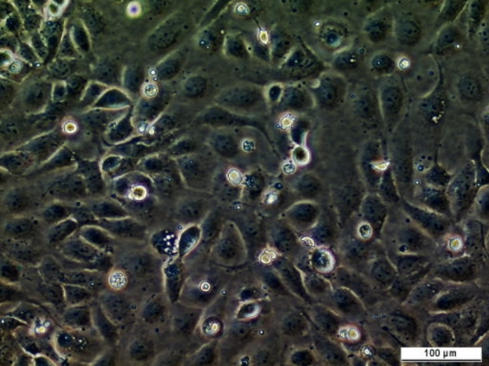
#161894

OCM.246

Please Enter Your Email ID
Email ID

Cat. #161894

OCM.246

Cat. #: 161894

Organism: Human

Tissue: Ovary. NOTE: The precursor of a substantial proportion of HGSOC is likely to be serous tubal intraepithelial carcinoma in the fimbriae of the fallopian tubes

£575.00

This fee is applicable only for non-profit organisations. If you are a for-profit organisation or a researcher working on commercially-sponsored academic research, you will need to contact our licensing team for a commercial use license.

Contributor

Inventor: Louisa Nelson and Stephen S. Taylor

Institute: University of Manchester

Primary Citation: Camilla Coulson-Gilmer et al. 2021. J Exp Clin Cancer Res. 16,40(1):323. PMID: 34656146

Tool Details
Applications
Handling
References

Tool Details

*FOR RESEARCH USE ONLY

  • Name: OCM.246
  • Cancer: Gynaecologic cancer
  • Research fields: Cancer
  • Organism: Human
  • Gender: Female
  • Tissue: Ovary. NOTE: The precursor of a substantial proportion of HGSOC is likely to be serous tubal intraepithelial carcinoma in the fimbriae of the fallopian tubes
  • Donor: Age at diagnosis: 62; Previously treated (4 lines); Germline BRCA2 mutation (c.5946del; [p.Ser1982ArgfsTer22]) Frameshift
  • Morphology: Epithelial
  • Growth properties: Adherent
  • Model description: FIGO 4A; Putative BRCA2 reversion mutation (c.5087_6841del [p.Ile1697_Gly2281del]; c.4946_6841del [p.Lys1649_Gly2281delinsArg]); HRP (RAD51 assay); TP53 mutation (nonsense)
  • Crispr: No
  • Description: The Taylor lab Ovarian Cancer Models (OCMs) are patient-derived tumour cell cultures, unfettered by contaminating, genetically normal stromal cells and the microenvironment. OCMs display the hallmark characteristics of HGSOC and retain the unique molecular features of the original tumour. OCMs have extensive proliferative potential, enabling high-resolution cell biology and drug-sensitivity profiling on tumour cells ‘close to the patient’, i.e. without extensive culture and genetic drift. OCMs are clinically annotated, and have been extensively characterised with associated data sets being publicly available (including multi-omics data e.g. exome and RNA sequencing, and single-cell karyotyping). OCMs can also be used to generate PDX, thus enabling in vivo studies of novel therapies. Collectively, this living biobank represents comprehensive platform for basic research and drug development projects (see PMID: 37592171 for a review)
  • Application: Ex vivo drug-sensitivity profiling; OCM-derived xenograft generation
  • Production details: To establish cultures, red blood cells were lysed, the remaining cellular fraction harvested by centrifugation, disaggregated if necessary then plated in OCMI media. Serial passaging and selective detachment eliminated white blood cells and yielded separate tumour and stromal fractions.
  • Biosafety level: 2

Applications

  • Application: Ex vivo drug-sensitivity profiling; OCM-derived xenograft generation

Handling

  • Format: Frozen
  • Passage number: Approx. P20
  • Growth medium: OCMI (PMID: 26080861; 32054838) + 5% hyclone FBS
  • Temperature: 37° C
  • Atmosphere: 5% CO2 & 5% O2
  • Shipping conditions: Dry ice
  • Storage medium: Bambanker freezing solution
  • Storage conditions: Liquid nitrogen (1/2 T75 ISO)
  • Initial handling information: Primaria™ Tissue Culture Flasks
  • Subculture routine: Split sub-confluent cultures (~90%) 1:2 using 0.25% trypsin. Limit trypsin incubation to 3 mins max, multiple applications of trypsin may be required to remove all the cells from the flask. Trypsin should be neutralised using DMEM containing 20% FBS
  • Cultured in antibiotics: Penicillin and Streptomycin
  • Mycoplasma free: Yes
  • Characterisation tests: TP53 mutation status confirmed by Sanger sequencing of RT-PCR products

References

  • Camilla Coulson-Gilmer et al. 2021. J Exp Clin Cancer Res. 16,40(1):323. PMID: 34656146