The biology of ovarian cancer can change dramatically as disease progresses, and treatment resistance evolves. Developed by Dr. Simon Langdon and colleagues at Cancer Research UK Scotland Centre, the PEO cell line series offers a unique longitudinal approach to studying these changes, with models representing different stages of disease and treatment response. Explore the story behind the PEO series, the distinct value of its models, and how they continue to support research into DNA repair, treatment resistance, and emerging therapeutic strategies.
Introduction
Despite decades of progress, ovarian cancer remains one of the most challenging cancers to study and treat. High-grade serous ovarian cancer (HGSOC), the most common and aggressive subtype, is characterised by extensive genomic instability, tumour heterogeneity and a high likelihood of relapse following initial treatment. While many patients respond well to platinum-based chemotherapy, resistance frequently develops, creating an ongoing need to understand disease progression and develop more effective therapeutic strategies.
To do this, we need clinically relevant models that capture the biological complexity of ovarian cancer and reflect how tumours evolve in response to treatment. Yet many traditional ovarian cancer cell lines represent only a snapshot in time, limiting their ability to model one of the disease’s greatest challenges: the emergence of treatment resistance.

Recognising the need for clinically relevant ovarian cancer models, Dr. Simon Langdon and colleagues at the Cancer Research UK Scotland Centre developed what has become an established ovarian cancer model system: the PEO cell line series. Because the models were derived from sequential samples collected from the same patient, the series provides a valuable opportunity to study changes associated with disease progression and treatment response.
Its well-characterised cell lines have supported research into BRCA biology, homologous recombination (HR) deficiency, platinum resistance and PARP inhibitor response, demonstrating the lasting value of models that capture disease in a biologically relevant context.
A longitudinal model of disease progression
The PEO series (PEO1, PEO4, and PEO6) was developed with a simple but powerful objective: to create a longitudinal model system that more accurately reflects treatment-associated tumour evolution in ovarian cancer.
The cell lines were initially developed to explore mechanisms and features of platinum resistance. However, they were then used to investigate hormonal and growth factor control in this disease and other regulatory features of ovarian cancer. These insights proved useful for later translational and eventual clinical studies.
— Dr. Simon Langdon
Rather than establishing a single cell line from one tumour sample, Dr. Langdon and colleagues developed a series of related cell lines from sequential samples collected from the same patient over the course of their clinical journey1. This approach captures different stages of disease progression, from treatment-sensitive disease to chemotherapy-resistant states, creating a unique resource for studying how ovarian cancer changes over time.
This represented a significant step forward for ovarian cancer research. Many existing models originate from different patients, introducing variation in genetic background and disease history that can make it difficult to distinguish treatment-associated changes from inter-patient differences. By maintaining a shared patient background across the series, the PEO panel provides a powerful system for researchers to identify treatment- and progression-associated changes while reducing the confounding effects of interpatient variability. What’s more, the panel allows researchers to compare sequential tumour derived models from the same patient across treatment-sensitive and resistant disease states, allowing to decipher the molecular changes associated with disease progression and therapeutic exposure.
Exploring the PEO panel
While the strength of the PEO cell lines lies in its value as a panel, each model offers distinct characteristics that make it a valuable research tool in its own right.
Longitudinal cell lines
The core longitudinal series comprises PEO1, PEO4 and PEO6, which were derived from the same patient at different stages of treatment and disease progression.
PEO1 cell line represents the treatment-sensitive stage of the series. Derived from ascitic fluid collected at first relapse following initial chemotherapy, PEO1 is cisplatin-sensitive and shows marked sensitivity to PARP inhibitors such as olaparib. The line is also BRCA2-deficient, resulting in impaired HR repair, making it a valuable model for studying DNA damage response and the relationship between BRCA2 dysfunction and therapeutic sensitivity1-3.
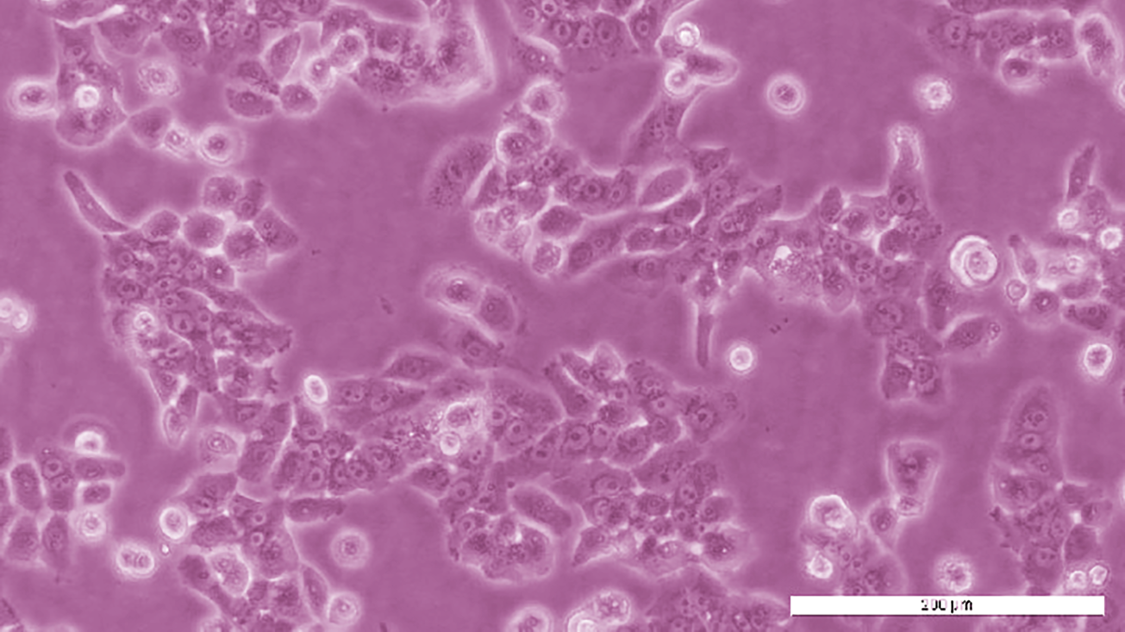
PEO4 cell line was established from the same patient following further disease progression and provides a complementary model of clinically acquired resistance and HR-restoration associated PARP inhibitor resistance. In contrast to PEO1, PEO4 has restored BRCA2 function through a secondary mutation, enabling recovery of HR repair capacity1,2,4. This paired relationship makes PEO1 and PEO4 particularly valuable for investigating how restoration of DNA repair can alter treatment response.
PEO6 represents a later treatment-resistant stage in the patient’s disease course. Like PEO4, PEO6 carries a secondary BRCA2 change that restores full-length BRCA and is considered HR-proficient. It is also resistant to cisplatin,1-4 providing researchers with an additional model for investigating tumour evolution and the biological changes associated with advanced disease.
We believe that these cell lines more accurately reflect the changes that can occur within the patient situation while undergoing treatment as opposed to changes developed in a cell-culture created context.
— Dr. Simon Langdon
Together, PEO1, PEO4 and PEO6 create a unique same-patient framework for researchers to track tumour evolution, treatment response, and acquired resistance.
In vitro resistant derivatives
Alongside the clinically derived longitudinal models, the PEO collection includes experimentally derived cell lines developed to investigate specific mechanisms of treatment resistance under controlled conditions.
PEO1-CDDP was developed through prolonged exposure of PEO1 cells to cisplatin, resulting in a model of acquired platinum resistance5. This provides a complementary system for investigating the cellular adaptations associated with sustained exposure to DNA-damaging therapy, supporting research into resistance mechanisms, biomarker discovery, and strategies to restore drug sensitivity.
PEO1-OR was developed from PEO1 through continuous, stepwise exposure to increasing concentrations of olaparib6. This model provides a system for studying acquired PARP inhibitor resistance and evaluating alternative therapeutic strategies in the context of olaparib-resistant disease.
Together, these derivatives extend the PEO toolkit by allowing researchers to compare resistance that emerged during the patient’s clinical course with resistance selected experimentally in vitro.
Additional patient-derived models
The PEO collection also includes additional cell lines that broaden the representation of patient-derived ovarian cancer biology beyond the original longitudinal series.
PEO14 and PEO23 were established from samples collected from the same patient at different stages of disease and treatment response. PEO23 represents a later platinum-resistant stage, providing a paired model system for investigating disease progression and the development of platinum resistance within a shared patient background. As such, PEO14 and PEO23 provide a complementary longitudinal perspective to the original PEO series and support research into the biological changes associated with treatment-resistant disease.
In addition to PEO14 and PEO23, there is the PEO16 cell line which was established from an independent patient sample. PEO16 provides an additional serous ovarian cancer model for comparative research, helping scientists to investigate ovarian cancer heterogeniety and explore how differences between tumours may influence disease biology and treatment response.
Together, the broader collection provides researchers with complementary models spanning disease progression, treatment sensitivity, acquired resistance, and DNA-repair biology.
From models to discoveries
The scientific value of the PEO series extends beyond its original development. As research questions have evolved, these models have continued to provide a foundation for investigating treatment response, resistance, and emerging therapeutic strategies.
A recent example comes from a 2026 study which investigated a combination of a checkpoint kinase 1 (CHK1) inhibitor with low-dose hydroxyurea as a potential treatment strategy for HGSOC8. The PEO1 was included as a core model within a broader HGSOC panel spanning different BRCA, HR-repair and treatment response backgrounds, with PEO4 providing an additional resistant, HR-restored comparator. The combination effectively induced cell death across the models tested, irrespective of BRCA2 and HR-repair status or prior chemotherapy response.
The researchers also investigated markers associated with immunogenic cell death. In PEO1, treatment significantly increased cell-surface HSP90 expression, demonstrating that the combination can trigger hallmarks of immunogenic cell death alongside its direct anti-tumour effects8.
The continued relevance of the PEO series can also be seen in research into platinum resistance. In a 2025 study, Adams et al. and colleagues evaluated the response of 36 ovarian cancer cell lines to cisplatin and carboplatin, including PEO1, PEO4 and PEO6 as a same-patient platinum-sensitive/resistant model set. The study used these models alongside other ovarian cancer cell lines to investigate genetic and gene-expression features associated with platinum response9.
Across the analysis, the researchers identified biological features associated with platinum resistance, including changes in innate immune/STAT signalling, EMT/WNT signalling, and regulators of platinum transport. The PEO series contributed directly to the study’s same patient comparison of platinum-sensitive and resistant HGSOC models, with PEO4 and PEO6 showing resistance-associated changes in pathways including innate inflammation/STAT signalling and platinum influx/efflux.
Such studies illustrate how the PEO panel continues to provide valuable reference points as researchers investigate the mechanisms underlying therapeutic resistance.
A lasting resource for ovarian cancer research
From its origins at the Cancer Research UK Scotland Centre in the late 1980s, to its continued use in modern research, the PEO series demonstrates the lasting value of researcher-developed cancer models. And as researchers seek robust, reproducible and translationally relevant systems, established models with a strong scientific foundation – such as the PEO panel – can provide an important starting point for new discoveries.
Given the ongoing use of platinum agents as first line therapy for ovarian cancer, we believe that these cell lines continue to be a relevant model of this disease to test new therapies and strategies.
— Dr. Simon Langdon
Three decades after the first PEO models were established, Dr. Langdon’s pioneering work continues to support research into disease biology, therapeutic resistance, and emerging approaches to treatment. Its story demonstrates how a researcher-developed innovation can extend far beyond its original purpose, providing a foundation for new questions as the field continues to evolve.
Explore the PEO cell line series to discover the models that could support your next study:
References
- Langdon et al. 1988. Cancer Research. 48(21):6166–6172. PMID: 3167863
- Sakai et al. 2009. Cancer Research. 69(16):6381–6386. PMID: 19654294
- Stukova et al. 2015. J Inorg Biochem. 149:45-8. PMID: 26021697
- Zhihong et al. 2016. Cancer Lett. 19;373(1):36–44. PMID: 26801746
- Macleod et al. 2005. Cancer Res. 65(15):6789-800. PMID: 16061661
- Biegala et al. 2023. Cells. 12(7), 1038. PMID: 37048111
- Langdon et al. 1990. Br J Cancer. 62(2):213-6. PMID: 2386737
- Zeng et al. 2026. Br J Cancer. 134(12):1830–40. PMID: 41946828
- Adams et al. 2025. Cancer Gene Ther. 32, 985–996. PMID:40683954