C32 Colorectal adenocarcinoma Cell Line
Exclusive toolA human colorectal adenocarcinoma model used to study colorectal cancer biology and therapeutic development. Exclusive to CancerTools. Save 30% on this cell line this autumn. Use code ACA26CELL at checkout.
Contributors
| Inventor | Institute |
|---|---|
| Walter Bodmer | University of Oxford |
| SKU: | 151756 |
|---|---|
| Product description: | This C32 cell line was established from a 66-year old female patient with a well differentiated adenocarcinoma of the colon classified as Dukes’ stage C. |
| Conditional: | Yes |
| Cellosaurus ID: | CVCL_5246 |
| Cat. #: | 151756 |
|---|---|
| Provenance and Ownership: | The C32 Colorectal adenocarcinoma Cell Line is owned by Cancer Research UK. CancerTools supplies the C32 Colorectal adenocarcinoma Cell Line under licence from Cancer Research UK and is the authorised source for its distribution. Sourcing C32 Colorectal adenocarcinoma Cell Line directly from the IP owner ensures authenticity, provenance and compliance with all applicable licensing and research use requirements. |
| Unit size: | 1×10^6 cells / vial |
| Cancer types: | Colorectal cancer |
| Research Fields: | Cancer;Drug development |
| Organism: | Human |
| Tissue: | Colon |
| Model: | Tumour line |
| Format: | Frozen |
|---|---|
| Shipping conditions: | Dry ice |
| Growth medium: | Iscove’s Modified Dulbecco’s Medium, + 10% Foetal Bovine Serum (FBS) + 2 mM Glutamine. |
| Subculture routine: | Split sub-confluent cultures (70-80%) 1:3 to 1:6 i.e. seeding at 2-4×10,000 cells/cm² using 0.05% trypsin or trypsin/EDTA; 5% CO₂; 37°C. C32 cells grow very slowly; following resuscitation or subculture the cells take at least 48 hours to re-attach. Cells should be left without disturbance during this time to facilitate adhesion. Centrifugation of the cells (100g x 5 min) at resuscitation to remove DMSO improves the establishment of a viable culture. Once attached, the cells grow in discrete islands and use of trypsin or trypsin/EDTA to subculture the cells (even without knocking the flask) yields large clumps. Further disaggregation may be achieved by repeatedly pipetting the cells. |
| References: |
Browning et al. 1993. Proc Natl Acad Sci U S A. 90(7):2842-5. PMID: 8464898. Tissue typing the HLA-A locus from genomic DNA by sequence-specific PCR: comparison of HLA genotype and surface expression on colorectal tumor cell lines. |
|---|
Images
View Gallery
Product Images
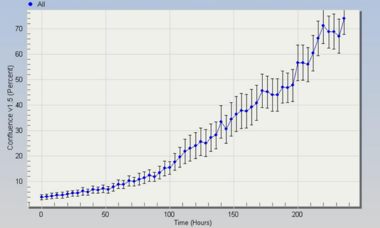
C32 Cell Line – growth profile.
C32 Cell Line – growth profile. Image courtesy of the European Collection of Authenticated Cell Cultures (ECACC), UK.
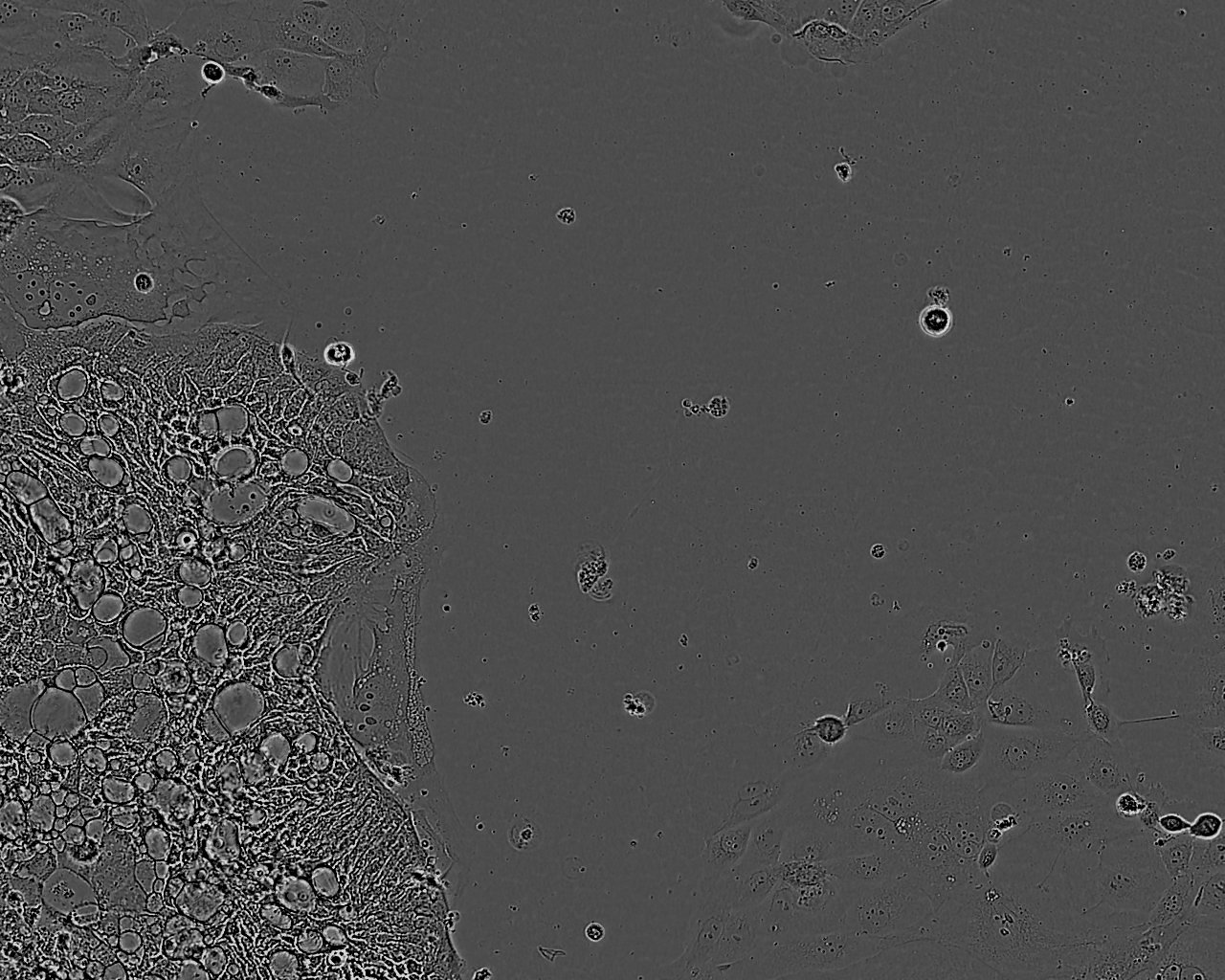
C32 Colorectal adenocarcinoma Cell Line. 6 days post plating. Image courtesy of the European Collection of Authenticated Cell Cultures (ECACC).