CD6-GFP Jurkat cell line
CD6 is a surface glycoprotein receptor, which has been previously shown to associate with CD3 and co-localize to the center of the IS in static conditions or stable T cell-APC contacts.
Contributors
| Inventor | Institute |
|---|---|
| Alessandra Cambi | Radboud University Medical Center |
| SKU: | 160714 |
|---|---|
| Product description: | Activation of the T cell receptor (TCR) on the T cell through ligation with antigen-MHC complex of an antigen-presenting cell (APC) is an essential process in the activation of T cells and induction of the subsequent adaptive immune response. Upon activation, the TCR, together with its associated co-receptor CD3 complex, assembles in signaling microclusters that are transported to the center of the organizational structure at the T cell-APC interface termed the immunological synapse (IS). During IS formation, local cell surface receptors and associated intracellular molecules are reorganized, ultimately creating the typical bull’s eye-shaped pattern of the IS. CD6 is a surface glycoprotein receptor, which has been previously shown to associate with CD3 and co-localize to the center of the IS in static conditions or stable T cell-APC contacts. |
| Alternate name: | CD6 |
| Conditional: | No |
| Production details: | Jurkat cell lines stably expressing CD6-GFP were obtained by electroporation using the Neon Transfection System for Electroporation (Invitrogen) according to the manufacturer’s guidelines. Shortly, 5*105 Jurkat cells were transfected at 1325 Volt (10 ms, 3 pulses) with 3 ÄÂÄ?g of DNA in 100 ÄÂÄ?l Resuspension buffer. After transfection cells were seeded in 2 ml of 1640 RPMI with 10% FCS and 1% U-glut. Antibiotics were added after 3 h. Stable cell lines were sorted on GFP expression on… |
| Parental cell line: | Jurkat E6.1 |
| Disease: | Cancer |
| Cat. #: | 160714 |
|---|---|
| Tool sub type: | Continuous |
| Unit size: | 1×10^6 cells / vial |
| Cancer types: | Blood cancer |
| Research Fields: | Cell biology; Cell signaling and signal transduction; Immunology |
| Organism: | Human |
| Tissue: | Blood |
| Cancer Types In Detail: | Acute T cell Leukemia |
| Format: | Frozen |
|---|---|
| Shipping conditions: | Dry ice |
| Growth medium: | 1640 RPMI supplemented with 10% Fetal Calf Serum, 1 mM Ultra-glutamine and antibiotics (100 U/ml penicillin, 100 ?g/ml streptomycin and 0.25 ?g/ml amphotericin B. 500 ng/ml geneticin. |
| References: |
Meddens et al. 2018. Front Immunol. 9:2333. PMID: 30356797. |
|---|
Images
View Gallery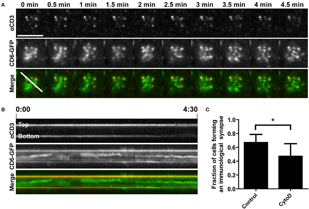
Product Images
Disruption of actin polymerization inhibits TCR/CD3 and CD6 co-transport toward the cSMAC of the immunological synapse on ?CD3-containing SLBs. (A;B) CD6-GFP Jurkat T cells; either untreated or pretreated with 0.5 ?M Cytochalasin D for 15 min; were seeded on a SLB containing ATTO647-conjugated ?CD3. Twenty minutes after seeding CD6-GFP and ?CD3 dynamics were imaged using TIRF microscopy live cell imaging. Snap shots of every 30 s of the time lapse are shown in (A). Kymograph of the line (indicated in the Merge image at time point 0 min in A) during the whole imaging period (4.5 min) is shown in (B). (C) Wildtype Jurkat T cells; either untreated or pretreated with 0.5 ?M Cytochalasin D for 15 min; were seeded for 30 min on SLBs containing ATTO647-conjugated ?CD3; and subsequently fixed. Widefield microscopy was performed and cells (>44 cells per condition; three independent experiments) were scored for synapse formation based on identification of cells by brightfield displaying ?CD3 positive cluster formation in a lipid bilayer (DiI) positive area. Average percentages of cells forming an immunological synapse are represented in (C). Bars represent mean with SD. Statistical significance was tested with paired t-test. Scale bar represents 10 ?m; *p < 0.05. Taken from Meddens MBM; Mennens SFB; Celikkol FB; Te Riet J; Kanger JS; Joosten B; Witsenburg JJ; Brock R; Figdor CG; Cambi A. Biophysical Characterization of CD6-TCR/CD3 Interplay in T Cells. Front Immunol. 2018 Oct 9;9