In conventional media, much of a cell’s trace element supply doesn’t come from the formulation at all, it comes from the serum, in amounts that vary from lot to lot. PlasmaxTM, developed by Dr. Saverio Tardito and his team at the Cancer Research UK Scotland Institute, takes a different approach. It specifies all six trace elements at defined concentrations in the base formulation, within a wider formulation calibrated to human plasma. Here’s what the evidence says about why that distinction matters.
A formulation gap worth knowing about
When selecting a cell culture medium, the conversation tends to focus on glucose concentration, glutamine levels, and serum supplementation. Trace elements are less often part of that discussion, despite playing essential roles in antioxidant defence, enzyme function, and cellular redox regulation.
Yet the trace element content of conventional media differs substantially from human plasma and, as the next section shows, a large part of what a cell receives doesn’t come from the base medium at all. The consequences are easiest to see in experiments that place real demand on a cell’s antioxidant capacity, such as low-density seeding and colony formation assays, or where oxidative stress is itself a variable of interest. Knowing what your medium contains, and where those components come from, is as important as any other experimental parameter.
What conventional media leave out
PlasmaxTM includes trace elements at concentrations calibrated to those found in human plasma: copper, iron, manganese, selenium, vanadium, and zinc1. RPMI 1640 contains none of them; DMEM/F-12 defines only copper, iron and zinc, and omits manganese, selenium and vanadium. Cells cultured in these media still receive trace elements, but mostly from the serum rather than the base formulation, which either omits them or supplies only part of what the cell needs. That distinction turns out to matter because trace elements are not just supplements; they are essential nutrients that can shape cell physiology and function.
Each element contributes to cellular function in a distinct way. Zinc (Zn) is a structural cofactor for hundreds of enzymes and contributes to gene regulation and antioxidant defence. Copper (Cu) is a cofactor for antioxidant enzymes including superoxide dismutase, which neutralises reactive oxygen species generated during normal metabolism. Iron and manganese both support redox balance and mitochondrial function. Selenium is built into selenoproteins as the amino acid selenocysteine, positioned at their active site, where it underpins the cell’s antioxidant defences – including the activity of glutathione peroxidase 4 (GPX4)1. Vanadium behaves as a phosphate analogue and can affect the activity of enzymes such as protein tyrosine phosphatases, though its role in mammalian cells is less well defined.
Several of these elements underpin antioxidant defence and redox balance, so their availability shapes how your cells cope with oxidative stress, most visibly in the assays you run routinely: low-density seeding, single-cell cloning, and colony formation.
Selenium: what the evidence shows
Selenium provides a clear example of how trace element availability influences cells in culture, and the evidence spans five decades. In 1976, McKeehan et al reported that selenium (Se) is essential for clonal growth of human diploid fibroblasts in media containing low concentrations of serum protein. They found that higher serum concentrations masked the requirement entirely, because serum is itself a source of selenium2. This work was among the key findings that led to selenium becoming a standard component of serum-free and chemically defined media formulations.
Vande Voorde et al. later examined selenium’s role in cancer cells and identified the underlying mechanism. Supplementing with 2.5% foetal bovine serum (FBS) to minimise the contribution of serum components, they found that breast cancer cells seeded at low density in selenium-deprived medium formed far fewer colonies. Without selenium, cells cannot maintain sufficient GPX4 activity to protect against lipid peroxidation, and the result is ferroptosis – iron-dependent cell death driven by the nutrient environment. Reintroducing sodium selenite restored GPX4 activity and colony-forming capacity1.
Covered by serum is not the same as controlled
Both studies above point to the same underlying fact: serum is itself a source of selenium. At 10% FBS, it likely supplies enough to mask any deficiency, which is why the subject of trace elements rarely come up in routine culture discussions. But it also means the total trace element content of your experiments isn’t fully specified by the base medium. It depends on what the serum contained, at what concentration, in each batch.
For a great deal of routine work, that goes unnoticed and causes no problem. It becomes consequential in two situations: when serum is reduced for reproducibility, to limit confounding growth factors, or when experiments place real demand on antioxidant capacity, such as low-density seeding or protocols where oxidative stress is a variable of interest.
The difference between DMEM/F-12 medium supplemented with FBS versus PlasmaxTM supplemented with FBS is shown in Figure 1.
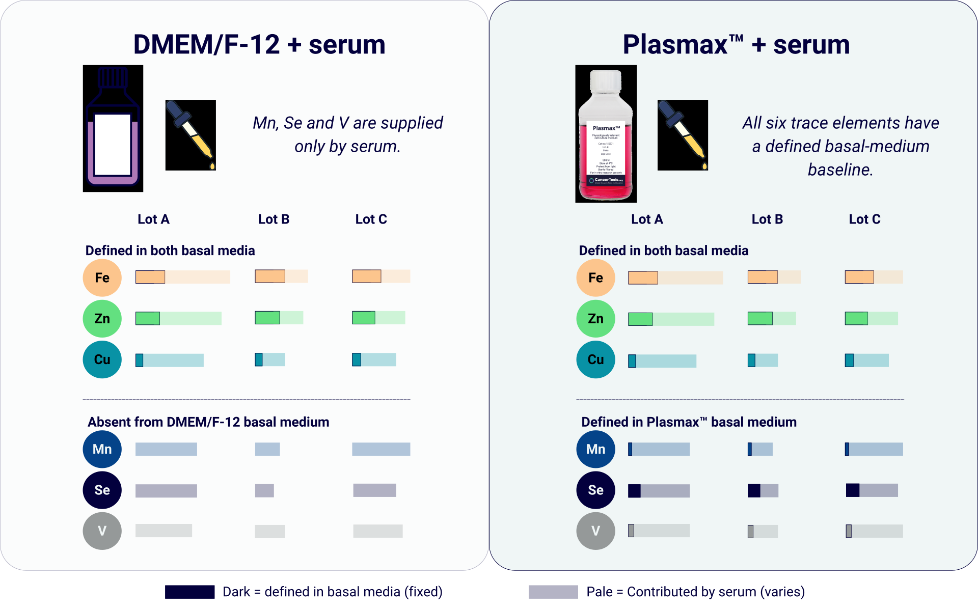
This shifts the relevant question from whether trace elements are present to whether their concentration is defined and reproducible.
How PlasmaxTM makes the input defined
PlasmaxTM supplies these trace elements at defined, physiologically relevant concentrations as part of the base formulation, not as variables contributed from serum. They sit within a wider formulation of 80 components calibrated to human plasma1.
The functional benefit extends beyond the selenium and ferroptosis mechanism. Golikov et al. found that cultivation in PlasmaxTM enhances mitochondrial respiratory capacity across four mammalian cell lines compared with conventional media3, evidence that a physiologically relevant formulation affects cellular function under standard conditions, beyond the low-serum, low-density context in which the ferroptosis effect was characterised.
The table below shows how PlasmaxTM compares across the formulation parameters that most affect biological fidelity and experimental outcomes.
Table 1. PlasmaxTM compared with conventional and physiologically relevant media across key formulation parameters.
| PlasmaxTM | Physiologically relevant competitor medium | DMEM/F-12 | RPMI 1640 | |
|---|---|---|---|---|
| Total no. of components | 80 | 74 | 52 | 40 |
| Defined trace elements in basal medium | Cu Fe Mn Se V Zn | Not specified | Cu Fe Zn | Not specified |
| FBS supplementation | 2.5 – 10%* | 10% | 10% | 10% |
| Physiological relevance1 | +++ | +++ | + | + |
| Supports growth at low density** | Demonstrated | Not evaluated | Lower than PlasmaxTM | Not evaluated |
** Based on Vande Voorde et al. (2019), which reported increased colony formation of three breast cancer cell lines seeded at low-density in PlasmaxTM compared to DMEM/F-12 under reduced-serum conditions (2.5% FBS), mediated by selenium-dependent GPX4 activity and protection from ferroptosis. Equivalent comparative studies were not identified for the other media shown1.
A variable worth controlling
Trace elements rarely make it onto a methods checklist. In conventional media they don’t need to – serum quietly covers the requirement, which is exactly why the variable goes unexamined. But covered is not the same as controlled, and the difference surfaces when serum is reduced or when an experiment places real demand on a cell’s antioxidant defences. PlasmaxTM specifies all six trace elements as part of the formulation, so the input is known rather than incidental.
Trace elements are one example of a broader principle. Because PlasmaxTM is calibrated to human plasma, its influence reaches well beyond antioxidant defence – from pyruvate-driven pseudohypoxic signalling to urea-cycle fidelity, and in bringing cultured cells’ metabolism closer to that of tumours in vivo. These effects are explored in Bridge the gap between in vivo and in vitro.
Science that keeps giving
PlasmaxTM is the product of years of work by Saverio Tardito and his team, who set out to make cell culture better reflect the physiology of human tumours. CancerTools, a non-profit platform, part of Cancer Research UK, makes that innovation accessible to researchers everywhere, extending its reach beyond a single lab and supporting more reproducible science across the field. Every tool accessed contributes directly to future cancer research through a model that supports both the inventing institution and Cancer Research UK. Choosing PlasmaxTM means your research investment goes further than the lab.
Further resources
References
- Vande Voorde et al. 2019. Sci Adv. 5(1):eaau7314. PMID: 30613774.
- McKeehan et al. 1976. Proc Natl Acad Sci USA. 73(6):2023-2027. PMID: 1064872.
- Golikov et al. 2022. Antioxidants. 11(1):97. PMID: 35052601.