What happens to tumour metabolism when standard-of-care treatment is viewed through a more physiological lens? Using PlasmaxTM, Maria Francesca Allega (University of Chicago, formally at Cancer Research UK Scotland Institute) and colleagues explored how dexamethasone (Dex) reshapes glioblastoma metabolism under conditions closer to human plasma. Their work uncovered a previously unrecognised steroid-driven metabolic rewiring centred on nicotinamide N-methyltransferase (NNMT) activity, nicotinamide metabolism, and methionine dependency – opening new avenues for metabolic imaging and intervention.
Introduction
Glioblastoma is the most common and aggressive malignant brain tumour in adults, with a median survival of just 12-15 months1. Despite intensive treatment, outcomes remain poor and the disease is characterised by profound biological and metabolic heterogeneity.
Importantly, glioblastoma-associated cerebral oedema is routinely managed using synthetic glucocorticoids such as Dexamethasone (Dex). While it is effective at reducing oedema and other neurological symptoms, the impact of these drugs on tumour metabolism has remained largely unexplored. Such unknowns represent ongoing challenges in the field and understanding treatment-induced metabolic changes requires robust experimental systems that can accurately reflect human physiology. However, conventional cell culture media often introduces non-physiological nutrient conditions that can distort metabolic phenotypes.
A recent study led by Maria Francesca Allega, Saverio Tardito and colleagues, published in Science Advances, set out to investigate whether glucocorticoid treatment directly alters glioblastoma metabolism and whether these effects could reveal new therapeutic or imaging opportunities2. The team adopted PlasmaxTM, a physiologically relevant plasma-like culture medium, to model tumour metabolism under nutrient conditions that more closely reflect human plasma. This approach allowed the team to identify a steroid-driven metabolic vulnerability linking nicotinamide and methionine metabolism.
“Dr. Saverio Tardito, my PhD supervisor, is the inventor of Plasmax™, so I was introduced to it as soon as I joined the lab. Because my project focused on tumour metabolism, it was a natural choice to perform the work in a physiological medium from the beginning. We wanted to study glioblastoma metabolism under conditions that better mimic human plasma rather than relying on conventional media known to impose metabolic artifacts.”
Maria Francesca Allega, PhD
Experimental approach
Allega and colleagues designed a multi-platform experimental workflow that combined physiologically relevant in vitro systems with in vivo validation.
Human glioblastoma models, including more than ten patient-derived, treatment-naïve cell lines, were cultured in serum-free PlasmaxTM from the outset. This provided a chemically defined, plasma-like nutrient environment with essential trace elements that supported cell growth without the need for foetal bovine serum (FBS), enabling a more controlled and reproducible study of metabolic activity under conditions closer to human physiology.
Cells were then treated with clinically relevant doses of Dex to model standard patient care. Metabolic responses were profiled using a combination of:
- Untargeted metabolomics to capture changes in intracellular and extracellular metabolites
- Stable isotope tracing (including labelled nicotinamide) to unravel pathway-level metabolic flux through key pathways
- Functional proliferation assays across multiple glioblastoma models to link metabolic changes to cellular behaviour
To determine whether these in vitro findings were maintained in vivo, orthotopic mouse models of glioblastoma were established by implanting tumour cells directly into the brain. Mice were treated with Dex, and tumour tissue was subsequently analysed using metabolomic profiling to validate whether the metabolic phenotype observed in vitro was preserved within the in vivo tumour microenvironment.
Results
Key findings from the study included:
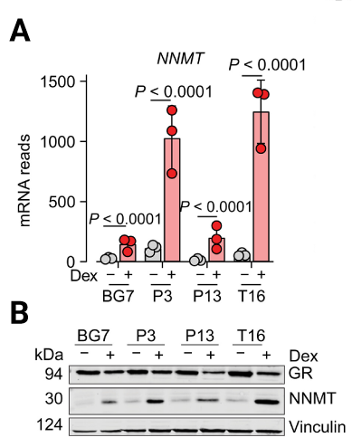
- Dex induces a distinct metabolic phenotype in glioblastoma: untargeted metabolic profiling showed that Dex treatment activated glucocorticoid receptor (GR) signalling and induced the accumulation of N1-methylnicotinamide, a product of nicotinamide N-methyltransferase (NNMT) activity, across multiple glioblastoma cells cultured in PlasmaxTM (Figure 1)
- Steroid treatment rewires nicotinamide metabolism in vitro: stable isotope tracing using labelled nicotinamide demonstrated that Dex redirected nicotinamide away from NAD+ biosynthesis and toward methylation, confirming activation of NNMT dependent metabolic rewiring.
- Methionine availability influences the metabolic response: Allega and colleagues found in vitro changes in methionine cycle intermediates, including increased demand for methyl donors such as S-adenosylmethionine (SAM). By modulating methionine levels under physiologically relevant conditions, the team showed that reduced methionine availability limited N1-methylnicotinamide accumulation following Dex treatment.
- The metabolic phenotype was validated in vivo: in orthotopic mouse models of glioblastoma, Dex treatment induced similar accumulation of N1-methylnicotinamide within tumour tissue, confirming that the metabolic rewiring observed in vitro was maintained in vivo.
- Glioblastoma cells displayed altered nutrient use patterns in vitro: under physiological nutrient conditions, glioblastoma cells preferentially consumed glutamate rather than glutamine, and in some cases, produced glutamine. This contrasts with the commonly reported “glutamine-addicted” phenotype observed in studies using conventional media.
- The findings enabled tumour-specific imaging in vivo: using radiolabelled nicotinamide in glioblastoma-bearing mice, the authors showed preferential accumulation of labelled nicotinamide within tumour regions using autoradiography and PET imaging. This signal reflected tumour-specific conversion of nicotinamide into N1-methylnicotinamide through NNMT activity and was enhanced following Dex treatment (Figure 3).
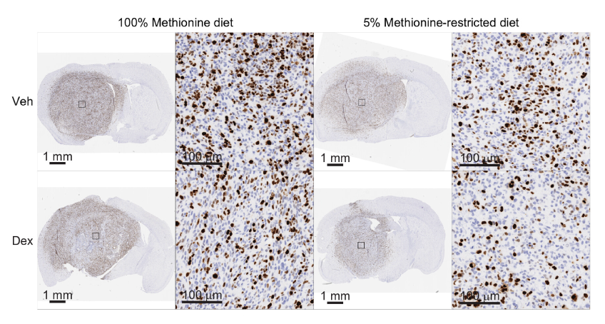
- Methionine restriction enhanced therapeutic response in vivo: combining Dex treatment with a methionine-restricted diet in the mouse models reduced methionine availability within tumour tissue and significantly decreased tumour cell proliferation compared with either treatment alone (Figure 2).
Discussion and impact
Dex induces a defined metabolic reprogramming in glioblastoma
This study demonstrates that Dex actively reshapes glioblastoma metabolism through NNMT-dependent rewiring of nicotinamide and methionine pathways. Rather than acting solely as a supportive therapy, Dex induces a reproducible metabolic phenotype characterised by increased conversion of nicotinamide to N1-methylnicotinamide.
In practical terms, this links a widely used clinical treatment to a specific and measurable metabolic signature, providing new insight into how standard therapies can directly influence tumour biology.
Nutrient availability shapes steroid-induced metabolic rewiring
Allega and colleagues further show that this metabolic shift is dependent on methionine availability, which provides the methyl donors required for NNMT-mediated conversion of nicotinamide to N1-methylnicotinamide.
By modulating methionine levels under physiologically relevant conditions, the team demonstrated that reduced methionine availability limits N1-methylnicotinamide accumulation following Dex treatment. Importantly, this dependency was confirmed in orthotopic mouse models, showing that the same metabolic relationship is maintained in vivo (Figure 2).
Together, these findings highlight a nutrient-sensitive vulnerability: as NNMT activity increases, glioblastoma cells become more dependent on methionine metabolism, creating a potential metabolic constraint that could be exploited therapeutically.
Physiological modelling reveals context-dependent metabolic behaviour
The study also identified metabolic behaviours that differ from commonly reported glioblastoma phenotypes. Under conditions that closely reflect human plasma, tumour cells preferentially consumed glutamate rather than glutamine, and in some cases produced glutamine, contrasting with the widely described “glutamine-addicted” phenotype observed in conventional culture systems.
This reinforces a key principle in cancer metabolism research: nutrient context strongly influences observed metabolic dependencies.
From metabolic rewiring to imaging and therapeutic opportunities
The accumulation of N1-methylnicotinamide in tumour tissue provides a clear biochemical signal of Dex-induced metabolic rewiring. To explore its translational potential, Allega and colleagues used radiolabelled nicotinamide in glioblastoma-bearing mice and demonstrated selective tumour accumulation using both autoradiography and PET imaging (Figure 3). This signal reflected tumour-specific conversion into N1-methylnicotinamide via NNMT activity and was enhanced following Dex treatment.

Because N1-methylnicotinamide carries a positive charge, it’s more readily retained within tumour cells, generating a detectable imaging signal. Importantly, the increased PET signal was not simply due to higher nicotinamide uptake but reflected tumour-specific metabolic rewiring. Together, these findings provide preclinical proof-of-principle for nicotinamide-based PET imaging, offering a non-invasive approach to visualise glioblastoma metabolism in vivo.
The role of physiological modelling in metabolic discovery
By using PlasmaxTM Allega and colleagues were able to study glioblastoma metabolism under nutrient conditions that more closely reflect human plasma. This reduced the risk that observed metabolic phenotypes were driven by non-physiological culture conditions, a known limitation of conventional media.
While PlasmaxTM does not fully reproduce the complexity of the tumour microenvironment, it provides a more physiologically grounded medium for studying nutrient-sensitive pathways. In this study, this approach supported findings that were consistent across in vitro models, orthotopic mouse systems, and patient-derived samples, strengthening their translational relevance.
“Plasmax™ gave us a serum-free, more physiologically relevant framework in which to study glioblastoma metabolism. It helped us interrogate the effects of dexamethasone under nutrient conditions closer to those experienced by cells in vivo, which increased our confidence in the biological relevance of the metabolic rewiring observed in cultured cells before validating it in vivo and in tumour samples from glioblastoma patients.”
Maria Francesca Allega, PhD
Conclusion
This study demonstrates how modelling tumour metabolism under physiologically relevant conditions can uncover clinically meaningful biology that may otherwise remain obscured in conventional systems. By using PlasmaxTM, Allega and colleagues identified a steroid-induced metabolic rewiring in glioblastoma that links Dex treatment to NNMT activity, methionine metabolism, and tumour-specific accumulation of N¹-methylnicotinamide – findings with direct implications for both metabolic imaging and therapeutic intervention.
Importantly, these discoveries translated beyond in vitro observations, remaining consistent across orthotopic mouse models and patient-derived samples. This reinforces the value of studying metabolism in nutrient environments that more closely reflect human physiology, particularly when investigating nutrient-sensitive pathways and metabolite exchange.
Looking ahead, the authors anticipate that PlasmaxTM will continue to play an important role in mechanistic and preclinical studies aimed at refining metabolic therapies and imaging strategies in glioblastoma and beyond.
“Because our work linked steroid treatment to a metabolic phenotype with direct imaging and therapeutic implications, I think Plasmax™ will remain very valuable in mechanistic studies and in preclinical work aimed at refining metabolic interventions.”
Maria Francesca Allega, PhD
For researchers seeking to generate more biologically relevant, reproducible, and translational metabolic data, PlasmaxTM offers a powerful platform to study cell metabolism under conditions closer to those encountered in vivo.
Explore how PlasmaxTM can support your metabolic research.
References
- Davis, M. 2016. Clinical Journal of Oncology Nursing. 20(5): S2–S8. PMID 27668386.
- Allega, M. F., et al., 2026. Science Advances. 12(4), eadx6539. PMID: 41576167.
