Melan-a Cell Line
Exclusive toolDiscover the melan-a cell line, an immortalised pigmented melanocyte line employed in the research of melanoma skin cancer, available exclusively at CancerTools.
Contributors
| Inventor | Institute |
|---|---|
| Dorothy Bennett | St George's University of London |
| SKU: | 153599 |
|---|---|
| Product description: | The melan-a cell line is an immortal melanocyte cell line derived from embryonic mouse skin. It was the first established non-tumorigenic mouse melanocyte cell line and has become a widely used model for melanocytes studies. Melanocytes are pigment-producing cells located in the bottom layer of the skin’s epidermis and in other tissues. The melan-a cells display the pigmentation and morphology typical of normal melanocytes, expressing both gp100 and the melanocyte marker melan-A. They are diploid in chromosome number and syngeneic with C57BL mice and B16 melanoma sublines, facilitating comparative investigations. These cells are visibly pigmented, smaller than B16 melanoma cells and retain all tested characteristics of normal melanocytes, except senescence. Among retained features, they also display a proliferative response to cholera toxin in the presence of tetradecanoyl phorbol acetate (TPA). The availability of robust cellular models that reflect normal skin melanocytes, such as the melan-a cell line, is essential for studying the etiology and abnormalities of skin cancers and melanomas, allowing comparisons at a molecular and cellular level. |
| Alternate name: | melan-a mouse melanocyte cell line; melan-a mouse cell line |
| CRISPR: | No |
| Conditional: | Yes |
| Production details: | Derived from normal epidermal melanoblasts from trunk skin of embryonic day 18 C57BL mice, a black or nonagouti (a/a) mouse |
| Cellosaurus ID: | CVCL_4624 |
| Parental cell line: | Mouse melanoblasts |
| Disease: | Cancer |
| Cat. #: | 153599 |
|---|---|
| Provenance and Ownership: | The Melan-a Cell Line is owned by Cancer Research UK. CancerTools supplies the Melan-a Cell Line under licence from Cancer Research UK and is the authorised source for its distribution. Sourcing Melan-a Cell Line directly from the IP owner ensures authenticity, provenance and compliance with all applicable licensing and research use requirements. |
| Tool sub type: | Continuous |
| Unit size: | 1×10^6 cells / vial |
| Cancer types: | Skin cancer |
| Organism: | Mouse |
| Tissue: | Embryonic skin |
| Model: | Immortalised line; Non-tumorigenic in syngeneic and nude mice |
| Cancer Types In Detail: | Melanoma |
| Primary citation: | Bennett et al. 1987. Int J Cancer. 39(3):414-8. PMID: 3102392 |
| Format: | Frozen |
|---|---|
| Storage conditions: | Liquid Nitrogen |
| Shipping conditions: | Dry ice |
| Growth medium: | RPMI 1640 gassed with 10% CO2 (pH 6.9-7.0), 2 mM glutamine, 10% FCS (Important: do not heat the serum, nor use heat-inactivated serum, as this destroys its stimulatory activity for melanocytes), and 200 nM TPA. Also requires tyrosinase inhibitor phenylthiourea (PTU) for freeze and thaw. Low pH is essential for pigmented melanocytes. If pH of stored medium rises noticeably (red or pink colour), re-gas from cylinder of 10% CO2 before use. Please see detailed notes in Product Datasheet in the Documentation tab. |
| Subculture routine: | Passage at ~80-85% confluency (doubling time ~36-48 hrs). Carefully remove the medium and rinse the flask or dish with 0.5 vol. of 1X PBS without CaCl2 and MgCl2 (PBSA), where 1 volume is the normal volume of medium for the vessel being sub-cultured, e.g. add 7.5 mL PBSA for a 15-mL flask. Aspirate after the rinse. Gently add 0.5 vol of 250 μg/mL trypsin and 200 μg/mL EDTA in PBSA. Quickly remove most of this, replacing enough to keep the surface wet (~0.2 vol.), and incubate in a 37°C incubator for 3-5 minutes. Inspect the culture by microscope and check for complete detachment of cells by gently rocking the culture. Do not tap or bang, but incubate further if needed, until detached (or maximum 10 min.) Add 0.5 vol. of cold growth medium (with 10% FCS, neutralizes trypsin) to the culture and wash/pipette up and down to make a single-cell suspension. Transfer to a suitable tube or round flask, e.g. 20 mL universal. Rinse culture vessel with further growth medium/FCS to make up 1 vol. total and add to the suspension. Swirl to mix evenly. Count the cell density using a haemocytometer(s), keeping suspension on ice while counting, to prevent attachment to the tube. Dilute, add mitogens and replate at ~1.5 x10⁴/mL. Change medium every 4 days. |
| Temperature: | 37° C |
| Atmosphere: | 10% CO2 |
| Mycoplasma free: | Yes |
| Biosafety level: | 1 |
| References: |
Gabel et al. 2024. Nat Commun. 15(959):1-14. PMID: 38302465. Yasuta et al. 2024. Sci Rep. 14:1525. PMID: 38233537. Unapanta et al. 2023. J Biol Chem. 299(10):105192. PMID: 37625589. Schadendorf et. al. 2018. Nat Rev Dis Primers. 392(10151): 971-984. PMID: 30238891 Bennett et al. 1987. Int J Cancer. 39(3):414-418. PMID: 3102392. |
|---|
Images
View Gallery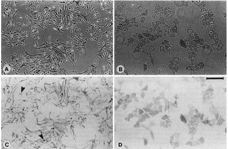
Product Images
 Powered by Bioz