Same cells. Different medium. Different biology.
Developed by Chris Halsey, Ekaterini Himonas, Vignir Helgason & Saverio Tardito at the University of Glasgow and the Cancer Research UK Scotland Institute, CSFmax is a cell culture medium modelled on human cerebrospinal fluid (CSF), designed to give scientists a more physiologically relevant model of the central nervous system (CNS) environment, starting with leukaemia.
Standard culture media such as RPMI 1640 contain about four times the key nutrients found in human cerebrospinal fluid, and nutrient levels in culture can change how cancer cells behave. CSFmax is a medium formulated from the nutrient concentrations of healthy human CSF, designed for researchers studying cells in or near the central nervous system (CNS), starting with CNS leukaemia. In a recent preprint, acute lymphoblastic leukaemia (ALL) cells grown in CSFmax grew more slowly and became sensitive to autophagy inhibition, a sensitivity not seen in standard media. If your cancer cell model migrates or metastasises to the CNS, a medium modelled on CSF may give more physiologically relevant answers.

Your medium is part of your experiment
Most culture media are designed to keep cells growing, not to mirror the fluid that surrounds them in the body. Published studies show that culture nutrient levels can rewire cancer cell metabolism. CSFmax is built from the mean metabolite concentrations reported for healthy human CSF in the Human Metabolome Database. As a result, its total nutrient concentration (≈ 4.2 mM) is much closer to human CSF (≈ 4.1 mM), compared to RPMI 1640 (~17.5 mM).
It exposed a dependency that standard media missed. In a recent preprint, B-cell acute lymphoblastic leukaemia (ALL) cell lines were grown in CSFmax, in standard RPMI 1640 and in a plasma-like medium and revealed:
- Slower growth, higher autophagy: cells in CSFmax proliferated more slowly than in either comparison medium and showed higher autophagy activity.
- A different metabolic state: compared with a plasma-like medium, SEM cells in CSFmax consumed fewer nutrients, had lower ATP and showed higher mitochondrial ROS. The authors compared these features with leukaemia cells isolated from the CNS of mouse xenografts.
- A dependency that standard media missed: a ULK1 inhibitor and bafilomycin A1 triggered cell death in CSFmax but not in the comparison media.
- Consistency in vivo: in mouse xenografts, SEM cells lacking ULK1 or ATG7 gave lower CNS leukaemia burden in three separate mouse xenograft experiments.
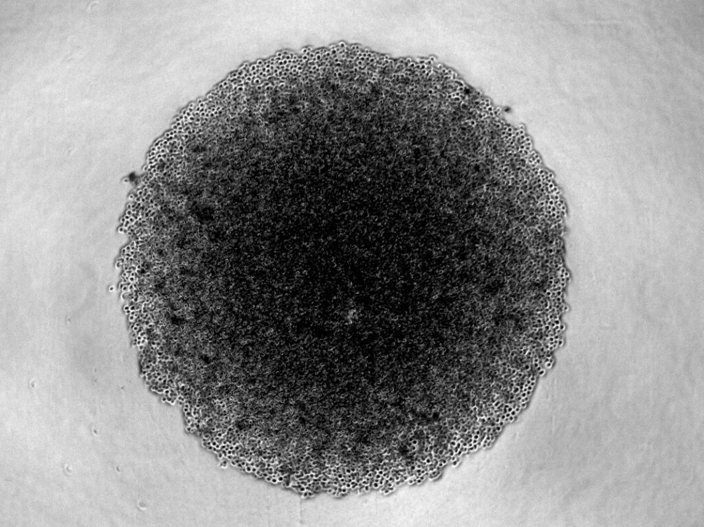
At a glance, CSFmax is:
- Modelled on human CSF: mean metabolite concentrations from healthy individuals (Human Metabolome Database).
- Close to CSF nutrient levels: ≈ 4.2 mM total nutrient concentration, versus about 4.1 mM in human CSF and about 17.5 mM in RPMI 1640.
- Tested so far: B-ALL cell lines SEM and REH in normoxic and hypoxic conditions in vitro and in vivo in xenograft studies.
Join the CSFmax waitlist
Exclusively available from CancerTools. Register your interest to be the first to receive launch details as soon as they’re announced.